Explore
 Validate
 Learn
Learn Western blot
Antibody data
- Antibody Data
- Antigen structure
- References [0]
- Comments [0]
- Validations
- Western blot [7]
- Immunocytochemistry [2]
- Immunoprecipitation [1]
- Other assay [1]
Submit
Validation data
Reference
Comment
Report error
- Product number
- PA5-85581 - Provider product page

- Provider
- Invitrogen Antibodies
- Product name
- VPS34 Polyclonal Antibody
- Antibody type
- Polyclonal
- Antigen
- Recombinant full-length protein
- Description
- Keep as concentrated solution. Predicted reactivity: Mouse (100%), Rat (100%), Zebrafish (86%), Xenopus laevis (92%), Dog (99%), Pig (98%), Bovine (99%). Positive Control: 293T, A431, HeLa, HepG2, U87-MG, SK-N-SH, IMR32, SK-N-AS, unboiled hVps34-transfected 293T cells. Store product as a concentrated solution. Centrifuge briefly prior to opening the vial.
- Reactivity
- Human
- Host
- Rabbit
- Isotype
- IgG
- Vial size
- 100 μL
- Concentration
- 1 mg/mL
- Storage
- Store at 4°C short term. For long term storage, store at -20°C, avoiding freeze/thaw cycles.
No comments: Submit comment
Supportive validation
- Submitted by
- Invitrogen Antibodies (provider)
- Main image
- Experimental details
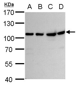
- VPS34 Polyclonal Antibody detects VPS34 protein by western blot analysis. A. 30 µg U87-MG whole cell lysate/extract. B. 30 µg SK-N-SH whole cell lysate/extract. C. 30 µg IMR32 whole cell lysate/extract. D. 30 µg SK-N-AS whole cell lysate/extract.7.5 % SDS-PAGE. VPS34 Polyclonal Antibody (Product # PA5-85581) dilution: 1:1,000.
- Submitted by
- Invitrogen Antibodies (provider)
- Main image
- Experimental details
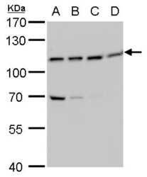
- VPS34 Polyclonal Antibody detects VPS34 protein by western blot analysis. A. 30 µg 293T whole cell lysate/extract. B. 30 µg A431 whole cell lysate/extract. C. 30 µg HeLa whole cell lysate/extract. D. 30 µg HepG2 whole cell lysate/extract.7.5 % SDS-PAGE. VPS34 Polyclonal Antibody (Product # PA5-85581) dilution: 1:1,000.

- Submitted by
- Invitrogen Antibodies (provider)
- Main image
- Experimental details
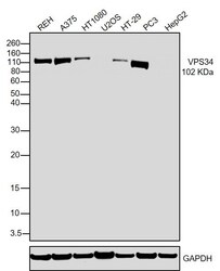
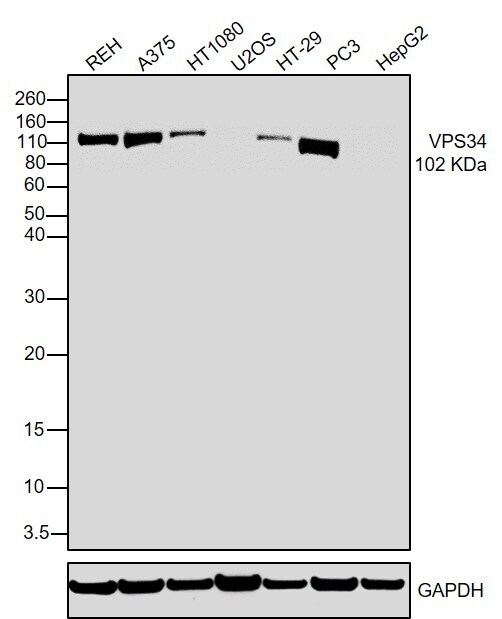
- Western blot was performed using Anti-VPS34 Polyclonal Antibody (Product # PA5-85581) and a 102 kDa band corresponding to Phosphatidylinositol 3-kinase catalytic subunit type 3 was observed across cell lines. Whole cell extracts (30 µg lysate) of Reh (Lane 1), A-375 (Lane 2), HT-1080 (Lane 3), U-2 OS (Lane 4), HT-29 (Lane 5), PC-3 (Lane 6), Hep G2 (Lane 7) were electrophoresed using NuPAGE™ 4-12% Bis-Tris Protein Gel (Product # NP0322BOX). Resolved proteins were then transferred onto a nitrocellulose membrane (Product # IB23001) by iBlot® 2 Dry Blotting System (Product # IB21001). The blot was probed with the primary antibody (1:1000) and detected by chemiluminescence with Goat anti-Rabbit IgG (Heavy Chain) Superclonal™ Recombinant Secondary Antibody, HRP (Product # A27036,1:4000) using the iBright™ FL1500 Imaging System (Product # A44115). Chemiluminescent detection was performed using SuperSignal™ West Pico PLUS Chemiluminescent Substrate (Product # 34580).
- Submitted by
- Invitrogen Antibodies (provider)
- Main image
- Experimental details
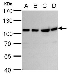
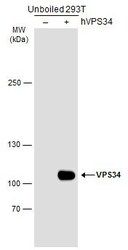
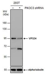
- Western Blot analysis of VPS34 was performed by separating 30 µg of non-transfected (–) and transfected (+) unboiled 293T whole cell extracts by 5% SDS-PAGE. Proteins were transferred to a membrane and probed with a VPS34 Polyclonal Antibody (Product # PA5-85581) at a dilution of 1:5000. The HRP-conjugated anti-rabbit IgG antibody was used to detect the primary antibody.
- Submitted by
- Invitrogen Antibodies (provider)
- Main image

- Experimental details
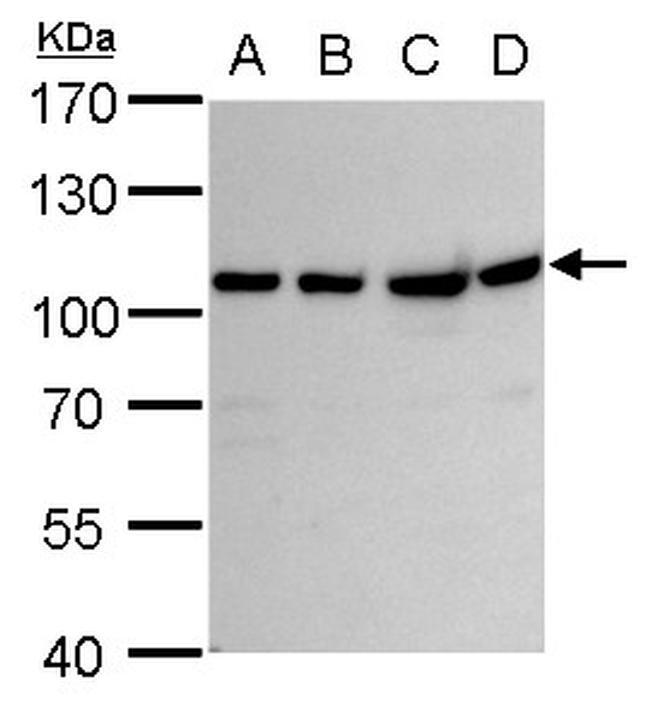
- Western blot analysis of VPS34 in A) U87-MG whole cell lysate, B) SK-N-SH whole cell lysate, C) IMR32 whole cell lysate, D) SK-N-AS whole cell lysate using VPS34 polyclonal antibody (Product # PA5-85581) using 30 µg of sample at a dilution of 1:1000. Prior to incubation with primary antibody, the sample was separated on 7.5% SDS-PAGE.
- Submitted by
- Invitrogen Antibodies (provider)
- Main image
- Experimental details
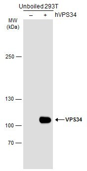
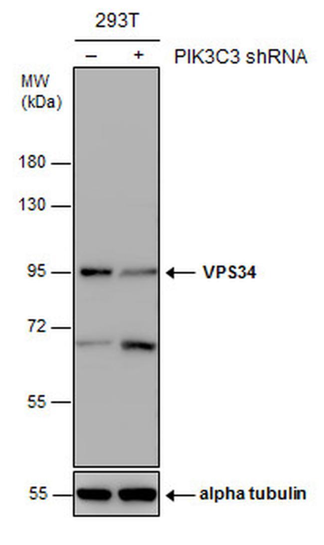
- Western blot analysis of VPS34 in non-transfected (–) and transfected (+) 293T whole cell extracts using VPS34 polyclonal antibody (Product # PA5-85581) using 30 µg of sample at a dilution of 1:1000. Prior to incubation with primary antibody, the sample was separated on 7.5% SDS-PAGE.
- Submitted by
- Invitrogen Antibodies (provider)
- Main image
- Experimental details
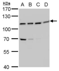
- Western blot analysis of VPS34 in A) 293T whole cell lysate, B) A431 whole cell lysate, C) HeLa whole cell lysate, D) HepG2 whole cell lysate using VPS34 polyclonal antibody (Product # PA5-85581) using 30 µg of sample at a dilution of 1:1000. Prior to incubation with primary antibody, the sample was separated on 7.5% SDS-PAGE.

Supportive validation
- Submitted by
- Invitrogen Antibodies (provider)
- Main image

- Experimental details
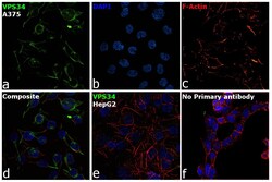
- Immunofluorescence analysis of Phosphatidylinositol 3-kinase catalytic subunit type 3 was performed using 70% confluent log phase A-375 cells. The cells were fixed with 4% paraformaldehyde for 10 minutes, permeabilized with 0.1% Triton™ X-100 for 15 minutes, and blocked with 2% BSA for 1 hour at room temperature. The cells were labeled with VPS34 Polyclonal Antibody (Product # PA5-85581) at 1:100 in 0.1% BSA, incubated at 4 degree celsius overnight and then labeled with Donkey anti-Rabbit IgG (H+L) Highly Cross-Adsorbed Secondary Antibody, Alexa Fluor Plus 488 (Product # A32790), (1:2000), for 45 minutes at room temperature (Panel a: Green). Nuclei (Panel b: Blue) were stained with ProLong™ Diamond Antifade Mountant with DAPI (Product # P36962). F-actin (Panel c: Red) was stained with Rhodamine Phalloidin (Product # R415, 1:300). Panel d represents the merged image showing Cytoplasmic localization. Panel e represents HepG2. Panel f represents control cells with no primary antibody to assess background. The images were captured at 60X magnification.

- Submitted by
- Invitrogen Antibodies (provider)
- Main image
- Experimental details
- Immunofluorescence analysis of Phosphatidylinositol 3-kinase catalytic subunit type 3 was performed using 70% confluent log phase A-375 cells. The cells were fixed with 4% paraformaldehyde for 10 minutes, permeabilized with 0.1% Triton™ X-100 for 15 minutes, and blocked with 2% BSA for 1 hour at room temperature. The cells were labeled with VPS34 Polyclonal Antibody (Product # PA5-85581) at 1:100 in 0.1% BSA, incubated at 4 degree celsius overnight and then labeled with Donkey anti-Rabbit IgG (H+L) Highly Cross-Adsorbed Secondary Antibody, Alexa Fluor Plus 488 (Product # A32790), (1:2000), for 45 minutes at room temperature (Panel a: Green). Nuclei (Panel b: Blue) were stained with ProLong™ Diamond Antifade Mountant with DAPI (Product # P36962). F-actin (Panel c: Red) was stained with Rhodamine Phalloidin (Product # R415, 1:300). Panel d represents the merged image showing Cytoplasmic localization. Panel e represents HepG2. Panel f represents control cells with no primary antibody to assess background. The images were captured at 60X magnification.
Supportive validation
- Submitted by
- Invitrogen Antibodies (provider)
- Main image
- Experimental details
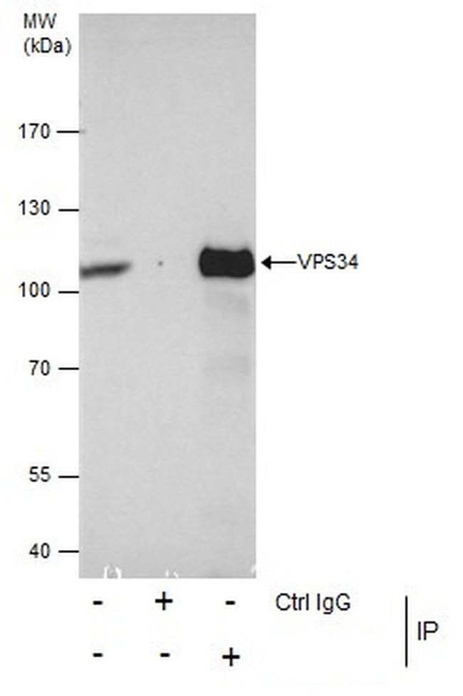
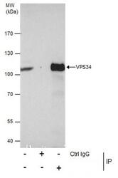
- Immunoprecipitation analysis of VPS34 in HeLa whole cell extracts with VPS34 polyclonal antibody (Product # PA5-85581) using 5 µg of sample, followed by anti-Rabbit IgG secondary antibody.

Supportive validation
- Submitted by
- Invitrogen Antibodies (provider)
- Main image

- Experimental details
- Immunoprecipitation analysis of VPS34 in HeLa whole cell extracts with VPS34 polyclonal antibody (Product # PA5-85581) using 5 µg of sample, followed by anti-Rabbit IgG secondary antibody.