Explore
 Validate
 Learn
Learn Western blot
 Immunocytochemistry
Antibody data
- Antibody Data
- Antigen structure
- References [1]
- Comments [0]
- Validations
- Immunocytochemistry [2]
- Immunohistochemistry [3]
- Other assay [2]
Submit
Validation data
Reference
Comment
Report error
- Product number
- MA5-29413 - Provider product page

- Provider
- Invitrogen Antibodies
- Product name
- CD146 Recombinant Rabbit Monoclonal Antibody (5)
- Antibody type
- Monoclonal
- Antigen
- Recombinant full-length protein
- Description
- This product is preservative free. It is recommended to add sodium azide to avoid contamination (final concentration 0.05%-0.1%). Recombinant rabbit monoclonal antibodies are produced using in vitro expression systems. The expression systems are developed by cloning in the specific antibody DNA sequences from immunoreactive rabbits. Then, individual clones are screened to select the best candidates for production. The advantages of using recombinant rabbit monoclonal antibodies include: better specificity and sensitivity, lot-to-lot consistency, animal origin-free formulations, and broader immunoreactivity to diverse targets due to larger rabbit immune repertoire. This antibody has specificity for Human CD146/MCAM.
- Reactivity
- Human
- Host
- Rabbit
- Isotype
- IgG
- Antibody clone number
- 5
- Vial size
- 100 μL
- Concentration
- 5 mg/mL
- Storage
- Store at 4°C short term. For long term storage, store at -20°C, avoiding freeze/thaw cycles.
Submitted references Wnt5a promotes renal tubular inflammation in diabetic nephropathy by binding to CD146 through noncanonical Wnt signaling.
Li X, Wen J, Dong Y, Zhang Q, Guan J, Liu F, Zhou T, Li Z, Fan Y, Wang N
Cell death & disease 2021 Jan 18;12(1):92
Cell death & disease 2021 Jan 18;12(1):92
No comments: Submit comment
Supportive validation
- Submitted by
- Invitrogen Antibodies (provider)
- Main image

- Experimental details
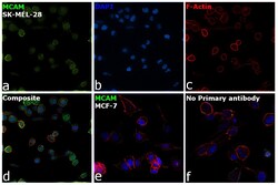
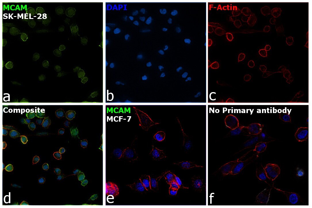
- Immunofluorescence analysis of Cell surface glycoprotein MUC18 was performed using 70% confluent log phase SK-MEL-28 cells. The cells were fixed with 4% paraformaldehyde for 10 minutes, permeabilized with 0.1% Triton™ X-100 for 15 minutes, and blocked with 2% BSA for 1 hour at room temperature. The cells were labeled with CD146 Recombinant Rabbit Monoclonal Antibody (5) (Product # MA5-29413) at 1:100 in 0.1% BSA, incubated at 4 degree celsius overnight and then labeled with Donkey anti-Rabbit IgG (H+L) Highly Cross-Adsorbed Secondary Antibody, Alexa Fluor Plus 488 (Product # A32790), (1:2000), for 45 minutes at room temperature (Panel a: Green). Nuclei (Panel b: Blue) were stained with ProLong™ Diamond Antifade Mountant with DAPI (Product # P36962). F-actin (Panel c: Red) was stained withRhodamine Phalloidin (Product # R415, 1:300). Panel d represents the merged image showing Membrane localization. Panel e represents Low expression in MCF-7. Panel f represents control cells with no primary antibody to assess background. The images were captured at 60X magnification.

- Submitted by
- Invitrogen Antibodies (provider)
- Main image
- Experimental details
- Immunofluorescence analysis of Cell surface glycoprotein MUC18 was performed using 70% confluent log phase SK-MEL-28 cells. The cells were fixed with 4% paraformaldehyde for 10 minutes, permeabilized with 0.1% Triton™ X-100 for 15 minutes, and blocked with 2% BSA for 1 hour at room temperature. The cells were labeled with CD146 Recombinant Rabbit Monoclonal Antibody (5) (Product # MA5-29413) at 1:100 in 0.1% BSA, incubated at 4 degree celsius overnight and then labeled with Donkey anti-Rabbit IgG (H+L) Highly Cross-Adsorbed Secondary Antibody, Alexa Fluor Plus 488 (Product # A32790), (1:2000), for 45 minutes at room temperature (Panel a: Green). Nuclei (Panel b: Blue) were stained with ProLong™ Diamond Antifade Mountant with DAPI (Product # P36962). F-actin (Panel c: Red) was stained withRhodamine Phalloidin (Product # R415, 1:300). Panel d represents the merged image showing Membrane localization. Panel e represents Low expression in MCF-7. Panel f represents control cells with no primary antibody to assess background. The images were captured at 60X magnification.
Supportive validation
- Submitted by
- Invitrogen Antibodies (provider)
- Main image

- Experimental details
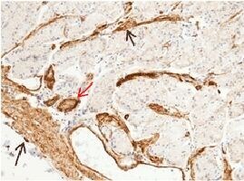
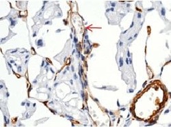
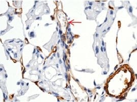
- Immunohistochemical staining of formalin fixed, paraffin-embedded human kidney showing membrane staining of endothelial cells (red arrow) using CD146 Recombinant Rabbit Monoclonal Antibody (5) (Product # MA5-29413, 1:1,000).
- Submitted by
- Invitrogen Antibodies (provider)
- Main image

- Experimental details
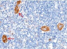
- Immunohistochemical staining of formalin fixed, paraffin-embedded human lymphonode showing membrane staining of endothelial cells (red arrow), smooth muscle cells (black arrow) using CD146 Recombinant Rabbit Monoclonal Antibody (5) (Product # MA5-29413, 1:1,000).
- Submitted by
- Invitrogen Antibodies (provider)
- Main image
- Experimental details
- Immunohistochemical staining of formalin fixed, paraffin-embedded human stomach showing membrane staining of endothelial cells (red arrow), smooth muscle cells (black arrow) using CD146 Recombinant Rabbit Monoclonal Antibody (5) (Product # MA5-29413, 1:1,000).
Supportive validation
- Submitted by
- Invitrogen Antibodies (provider)
- Main image
- Experimental details
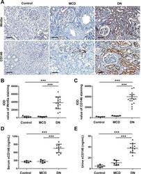
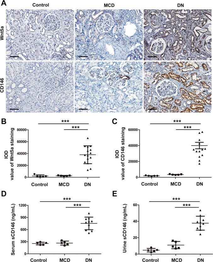
- Fig. 1 The expression levels of Wnt5a and CD146 were upregulated in diabetic neuropathy (DN) patients. A Representative immunostaining of Wnt5a and CD146 on normal kidney sections of nephrectomy samples ( n = 5) and kidney biopsy samples of patients with minimal change disease (MCD) ( n = 5) and DN ( n = 15). Original magnification, x200; scale bar, 100 mum. B Quantitative analysis of Wnt5a staining in kidney sections from each group according to the average integrated optical density (IOD). *** P < 0.001. C Quantitative analysis of expression of CD146 staining on kidney sections of each group by the average IOD. *** P < 0.001. D , E The concentration of soluble CD146 (sCD146) in serum and urine samples of each group. *** P < 0.001. Immunostaining was performed in duplicate. Significant differences were determined by unpaired two-tailed t -tests.
- Submitted by
- Invitrogen Antibodies (provider)
- Main image
- Experimental details
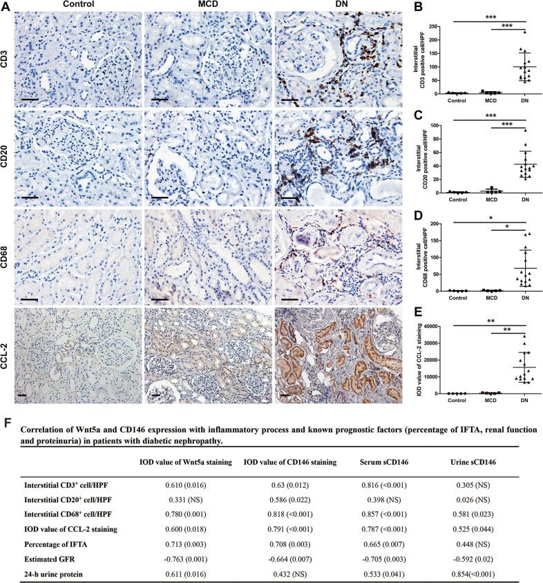
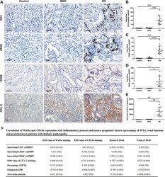
- Fig. 2 Assessment of renal inflammation in human kidney biopsies. A Immunostaining using specific antibodies for different immune cells, including T lymphocytes (CD3 + cells), B lymphocytes (CD20 + cells) and monocytes/macrophages (CD68 + cells), was performed on normal subjects, MCD patients, and DN patients. Original magnification, x400; scale bar, 50 mum. Immunohistochemical staining of the proinflammatory marker, chemokine (C-C motif) ligand 2 (CCL-2) was also performed. Original magnification, x200; scale bar, 50 mum. B - D The numbers of CD3 + , CD20 + , and CD68 + cells per high-power field (HPF) were counted in the tubulointerstitium. *** P < 0.001, * P < 0.05. E Quantitative analysis of the expression of CCL-2 staining on kidney sections of each group according to the average IOD. ** P < 0.01. Immunostaining was performed in duplicate. Significant differences were determined by unpaired two-tailed t -tests. F Correlation of Wnt5a and CD146 expression with inflammatory process and known prognostic factors (percentage of IFTA, renal function, and proteinuria) in patients with diabetic nephropathy. Data are r value based on Pearson and Spearman correlation analysis; ( P value); NS is not significant. IFTA, interstitial fibrosis and tubular atrophy; Estimated GFR, Estimated glomerular filtration rate; HbA1c, hemoglobin A1c.