 Explore
 Validate
 Learn
Learn Western blot
Antibody data
- Antibody Data
- Antigen structure
- References [6]
- Comments [0]
- Validations
- Western blot [2]
- Immunocytochemistry [1]
- Immunohistochemistry [3]
Submit
Validation data
Reference
Comment
Report error
- Product number
- AF932 - Provider product page

- Provider
- R&D Systems
- Product name
- Human MCAM/CD146 Antibody
- Antibody type
- Polyclonal
- Description
- Antigen Affinity-purified. Detects human MCAM/CD46 in Western blots and direct ELISAs. In direct ELISAs, less than 10% cross-reactivity with recombinant human (rh) ALCAM, rhBCAM, rhNCAM-L1, rhTROP-2 and recombinant mouse MCAM is observed.
- Reactivity
- Human
- Host
- Goat
- Conjugate
- Unconjugated
- Antigen sequence
AAA20824- Isotype
- IgG
- Vial size
- 100 ug
- Concentration
- LYOPH
- Storage
- Use a manual defrost freezer and avoid repeated freeze-thaw cycles. 12 months from date of receipt, -20 to -70 °C as supplied. 1 month, 2 to 8 °C under sterile conditions after reconstitution. 6 months, -20 to -70 °C under sterile conditions after reconstitution.
Submitted references Platelet-Derived Growth Factor Receptor-Positive Pericytic Cells of White Adipose Tissue from Critical Limb Ischemia Patients Display Mesenchymal Stem Cell-Like Properties.
Chemotherapy enhances tumor vascularization via Notch signaling-mediated formation of tumor-derived endothelium in breast cancer.
Expression of the immunoglobulin superfamily cell adhesion molecules in the developing spinal cord and dorsal root ganglion.
CD105 expression on CD34-negative spindle-shaped stromal cells of primary tumor is an unfavorable prognostic marker in early breast cancer patients.
Plasticity of blood- and lymphatic endothelial cells and marker identification.
Endothelial endoglin is involved in inflammation: role in leukocyte adhesion and transmigration.
Kim EJ, Seo SG, Shin HS, Lee DJ, Kim JH, Lee DY
Clinics in orthopedic surgery 2017 Jun;9(2):239-248
Clinics in orthopedic surgery 2017 Jun;9(2):239-248
Chemotherapy enhances tumor vascularization via Notch signaling-mediated formation of tumor-derived endothelium in breast cancer.
Zhang P, He D, Chen Z, Pan Q, Du F, Zang X, Wang Y, Tang C, Li H, Lu H, Yao X, Jin J, Ma X
Biochemical pharmacology 2016 Oct 15;118:18-30
Biochemical pharmacology 2016 Oct 15;118:18-30
Expression of the immunoglobulin superfamily cell adhesion molecules in the developing spinal cord and dorsal root ganglion.
Gu Z, Imai F, Kim IJ, Fujita H, Katayama Ki, Mori K, Yoshihara Y, Yoshida Y
PloS one 2015;10(3):e0121550
PloS one 2015;10(3):e0121550
CD105 expression on CD34-negative spindle-shaped stromal cells of primary tumor is an unfavorable prognostic marker in early breast cancer patients.
Martinez LM, Labovsky V, Calcagno ML, Davies KM, Garcia Rivello H, Bianchi MS, Wernicke A, Fernández Vallone VB, Chasseing NA
PloS one 2015;10(3):e0121421
PloS one 2015;10(3):e0121421
Plasticity of blood- and lymphatic endothelial cells and marker identification.
Keuschnigg J, Karinen S, Auvinen K, Irjala H, Mpindi JP, Kallioniemi O, Hautaniemi S, Jalkanen S, Salmi M
PloS one 2013;8(9):e74293
PloS one 2013;8(9):e74293
Endothelial endoglin is involved in inflammation: role in leukocyte adhesion and transmigration.
Rossi E, Sanz-Rodriguez F, Eleno N, Düwell A, Blanco FJ, Langa C, Botella LM, Cabañas C, Lopez-Novoa JM, Bernabeu C
Blood 2013 Jan 10;121(2):403-15
Blood 2013 Jan 10;121(2):403-15
No comments: Submit comment
Supportive validation
- Submitted by
- R&D Systems (provider)
- Main image
- Experimental details
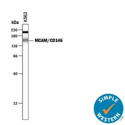
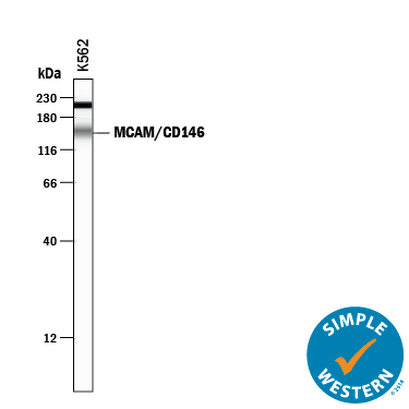
- Detection of Human MCAM/CD146 by Simple Western<sup abp="263">TM. Simple Western lane view shows lysates of K562 human chronic myelogenous leukemia cell line, loaded at 0.2 mg/mL. A specific band was detected for MCAM/CD146 at approximately 152 kDa (as indicated) using 10 µg/mL of Goat Anti-Human MCAM/CD146 Antigen Affinity-purified Polyclonal Antibody (Catalog # AF932) followed by 1:50 dilution of HRP-conjugated Anti-Goat IgG Secondary Antibody (Catalog # HAF109). This experiment was conducted under reducing conditions and using the 12-230 kDa separation system. Non-specific interaction with the 230 kDa Simple Western standard may be seen with this antibody.
- Submitted by
- R&D Systems (provider)
- Main image
- Experimental details
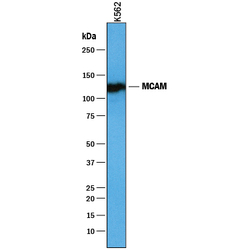
- Detection of Human MCAM/CD146 by Western Blot. Western blot shows lysates of K562 human chronic myelogenous leukemia cell line. PVDF membrane was probed with 1 µg/mL of Goat Anti-Human MCAM/CD146 Antigen Affinity-purified Polyclonal Antibody (Catalog # AF932) followed by HRP-conjugated Anti-Goat IgG Secondary Antibody (Catalog # HAF019). A specific band was detected for MCAM/CD146 at approximately 117 kDa (as indicated). This experiment was conducted under reducing conditions and using Immunoblot Buffer Group 1.

Supportive validation
- Submitted by
- R&D Systems (provider)
- Main image

- Experimental details
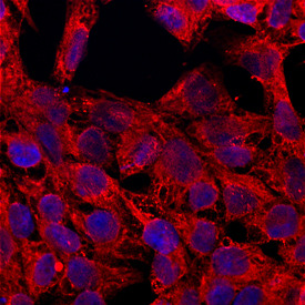
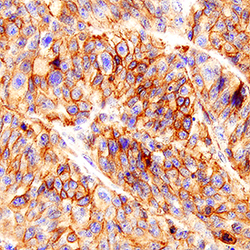
- MCAM/CD146 in BG01V Human Embryonic Stem Cells. MCAM/CD146 was detected in immersion fixed BG01V human embryonic stem cells using Goat Anti-Human MCAM/CD146 Antigen Affinity-purified Polyclonal Antibody (Catalog # AF932) at 10 µg/mL for 3 hours at room temperature. Cells were stained using the Northern-Lights™ 557-conjugated Anti-Goat IgG Secondary Antibody (red; Catalog # NL001) and counterstained with DAPI (blue). Specific staining was localized to cell membranes and cytoplasm. View our protocol for Fluorescent ICC Staining of Stem Cells on Coverslips.
Supportive validation
- Submitted by
- R&D Systems (provider)
- Main image

- Experimental details
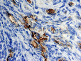
- MCAM/CD146 in Human Melanoma. MCAM/CD146 was detected in immersion fixed paraffin-embedded sections of human melanoma tissue using Goat Anti-Human MCAM/CD146 Antigen Affinity-purified Polyclonal Antibody (Catalog # AF932) at 15 µg/mL overnight at 4 °C. Tissue was stained using the Anti-Goat HRP-DAB Cell & Tissue Staining Kit (brown; Catalog # CTS008) and counterstained with hematoxylin (blue). View our protocol for Chromogenic IHC Staining of Paraffin-embedded Tissue Sections.
- Submitted by
- R&D Systems (provider)
- Main image
- Experimental details
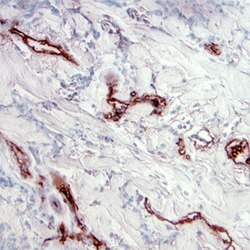
- MCAM/CD146 in Human Melanoma. MCAM/CD146 was detected in immersion fixed paraffin-embedded sections of human melanoma tissue using Goat Anti-Human MCAM/CD146 Antigen Affinity-purified Polyclonal Antibody (Catalog # AF932) at 5 µg/mL overnight at 4 °C. Tissue was stained using the Anti-Goat HRP-DAB Cell & Tissue Staining Kit (brown; Catalog # CTS008) and counterstained with hematoxylin (blue). View our protocol for Chromogenic IHC Staining of Paraffin-embedded Tissue Sections.
- Submitted by
- R&D Systems (provider)
- Main image
- Experimental details
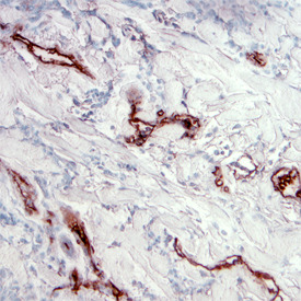
- MCAM/CD146 in Human Melanoma Tissue. MCAM/CD146 was detected in immersion fixed paraffin-embedded sections of human melanoma tissue using Goat Anti-Human MCAM/CD146 Antigen Affinity-purified Polyclonal Antibody (Catalog # AF932) at 3 µg/mL for 1 hour at room temperature followed by incubation with the Anti-Goat IgG VisUCyte™ HRP Polymer Antibody (Catalog # VC004). Tissue was stained using DAB (brown) and counterstained with hematoxylin (blue). Specific staining was localized to plasma membrane in cancer cells. View our protocol for IHC Staining with VisUCyte HRP Polymer Detection Reagents.
