Explore
 Validate
 Learn
Learn Flow cytometry
Antibody data
- Antibody Data
- Antigen structure
- References [5]
- Comments [0]
- Validations
- Flow cytometry [1]
- Other assay [2]
Submit
Validation data
Reference
Comment
Report error
- Product number
- 11-0748-42 - Provider product page

- Provider
- Invitrogen Antibodies
- Product name
- CD74 Monoclonal Antibody (5-329), FITC, eBioscience™
- Antibody type
- Monoclonal
- Antigen
- Other
- Description
- Description: This 5-329 monoclonal antibody recognizes human CD74, also known as the MHC Class II-associated invariant chain. A 33-43 kDa nonpolymorphic type II membrane protein, CD74 is highly expressed on B cells and subsets of activated T cells, Langerhans cells, dendritic cells, and epithelial cells. Monocytes and macrophages also express CD74, but at lower levels. CD74 acts as a chaperone of MHC Class II (HLA-DR) proteins, promoting their translocation from the ER to endocytic compartments during antigen presentation. Activation of NFkappaB and ERK has been shown following CD74 interaction with CD44 and binding to the proinflammatory cytokine macrophage migration-inhibitory factor (MIF). CD74 also plays a role in the maturation of follicular B-cells and accumulation of marginal zone B-cells. CD74 has also been demonstrated to interact with CXCR2 and CXCR4. Applications Reported: This 5-329 antibody has been reported for use in flow cytometric analysis. Applications Tested: This 5-329 antibody has been pre-titrated and tested by flow cytometric analysis of normal human peripheral blood cells. This can be used at 5 µL (0.125 µg) per test. A test is defined as the amount (µg) of antibody that will stain a cell sample in a final volume of 100 µL. Cell number should be determined empirically but can range from 10^5 to 10^8 cells/test. Excitation: 488 nm; Emission: 520 nm; Laser: Blue Laser. Filtration: 0.2 µm post-manufacturing filtered.
- Reactivity
- Human
- Host
- Mouse
- Conjugate
- Green dye
- Isotype
- IgG
- Antibody clone number
- 5-329
- Vial size
- 100 Tests
- Concentration
- 5 µL/Test
- Storage
- 4° C, store in dark, DO NOT FREEZE!
Submitted references Structural Remodeling of the Human Colonic Mesenchyme in Inflammatory Bowel Disease.
FOXP1 suppresses immune response signatures and MHC class II expression in activated B-cell-like diffuse large B-cell lymphomas.
A functional heteromeric MIF receptor formed by CD74 and CXCR4.
CD74 is a member of the regulated intramembrane proteolysis-processed protein family.
Invariant chain targets HLA class II molecules to acidic endosomes containing internalized influenza virus.
Kinchen J, Chen HH, Parikh K, Antanaviciute A, Jagielowicz M, Fawkner-Corbett D, Ashley N, Cubitt L, Mellado-Gomez E, Attar M, Sharma E, Wills Q, Bowden R, Richter FC, Ahern D, Puri KD, Henault J, Gervais F, Koohy H, Simmons A
Cell 2018 Oct 4;175(2):372-386.e17
Cell 2018 Oct 4;175(2):372-386.e17
FOXP1 suppresses immune response signatures and MHC class II expression in activated B-cell-like diffuse large B-cell lymphomas.
Brown PJ, Wong KK, Felce SL, Lyne L, Spearman H, Soilleux EJ, Pedersen LM, Møller MB, Green TM, Gascoyne DM, Banham AH
Leukemia 2016 Mar;30(3):605-16
Leukemia 2016 Mar;30(3):605-16
A functional heteromeric MIF receptor formed by CD74 and CXCR4.
Schwartz V, Lue H, Kraemer S, Korbiel J, Krohn R, Ohl K, Bucala R, Weber C, Bernhagen J
FEBS letters 2009 Sep 3;583(17):2749-57
FEBS letters 2009 Sep 3;583(17):2749-57
CD74 is a member of the regulated intramembrane proteolysis-processed protein family.
Becker-Herman S, Arie G, Medvedovsky H, Kerem A, Shachar I
Molecular biology of the cell 2005 Nov;16(11):5061-9
Molecular biology of the cell 2005 Nov;16(11):5061-9
Invariant chain targets HLA class II molecules to acidic endosomes containing internalized influenza virus.
Lamb CA, Yewdell JW, Bennink JR, Cresswell P
Proceedings of the National Academy of Sciences of the United States of America 1991 Jul 15;88(14):5998-6002
Proceedings of the National Academy of Sciences of the United States of America 1991 Jul 15;88(14):5998-6002
No comments: Submit comment
Supportive validation
- Submitted by
- Invitrogen Antibodies (provider)
- Main image
- Experimental details
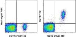
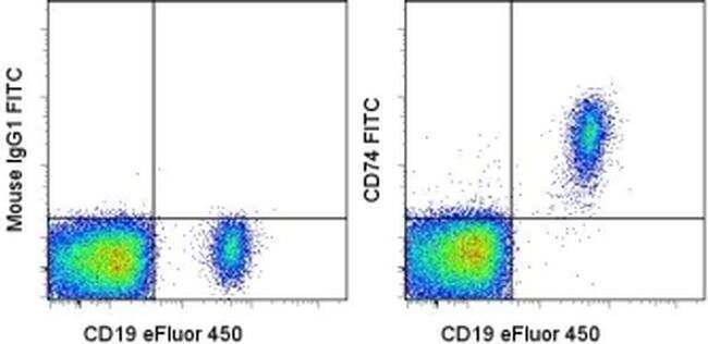
- Staining of normal human peripheral blood cells with Anti-Human CD19 eFluor® 450 (Product # 48-0199-42) and Mouse IgG1 K Isotype Control FITC (Product # 11-4714-42) (left) or Anti-Human CD74 FITC (right). Cells in the lymphocyte gate were used for analysis.
- Conjugate
- Green dye
Supportive validation
- Submitted by
- Invitrogen Antibodies (provider)
- Main image
- Experimental details
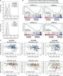
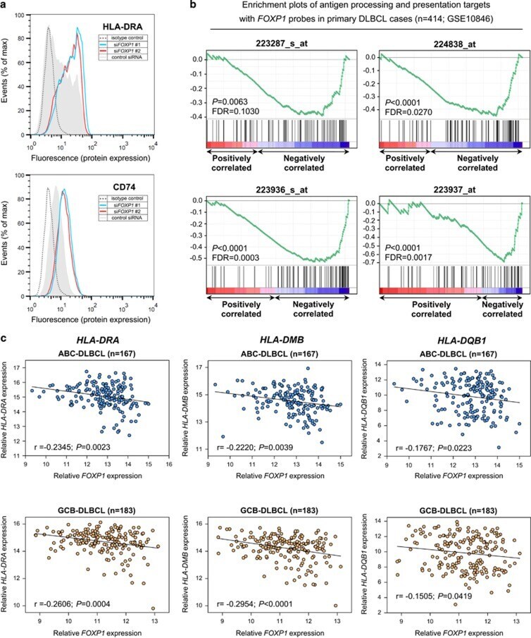
- Figure 4 FOXP1 silencing increases HLA-DRA expression in ABC-DLBCL while FOXP1 transcripts are inversely correlated with antigen processing/presentation and with individual MHC II genes in primary DLBCL. ( a ) Knockdown of FOXP1 in OCI-Ly3 cells increased HLA-DRA and CD74 protein expression on the cell surface. Flow cytometry plots shown are representative of three independent experiments. ( b ) Gene Set Enrichment Analysis of primary DLBCL cases ( n =414; GSE10846) for gene sets associated with FOXP1 transcript expression; the 'antigen processing and presentation' signature was significantly enriched according to four independent FOXP1 probes ( P
- Conjugate
- Green dye
- Submitted by
- Invitrogen Antibodies (provider)
- Main image
- Experimental details
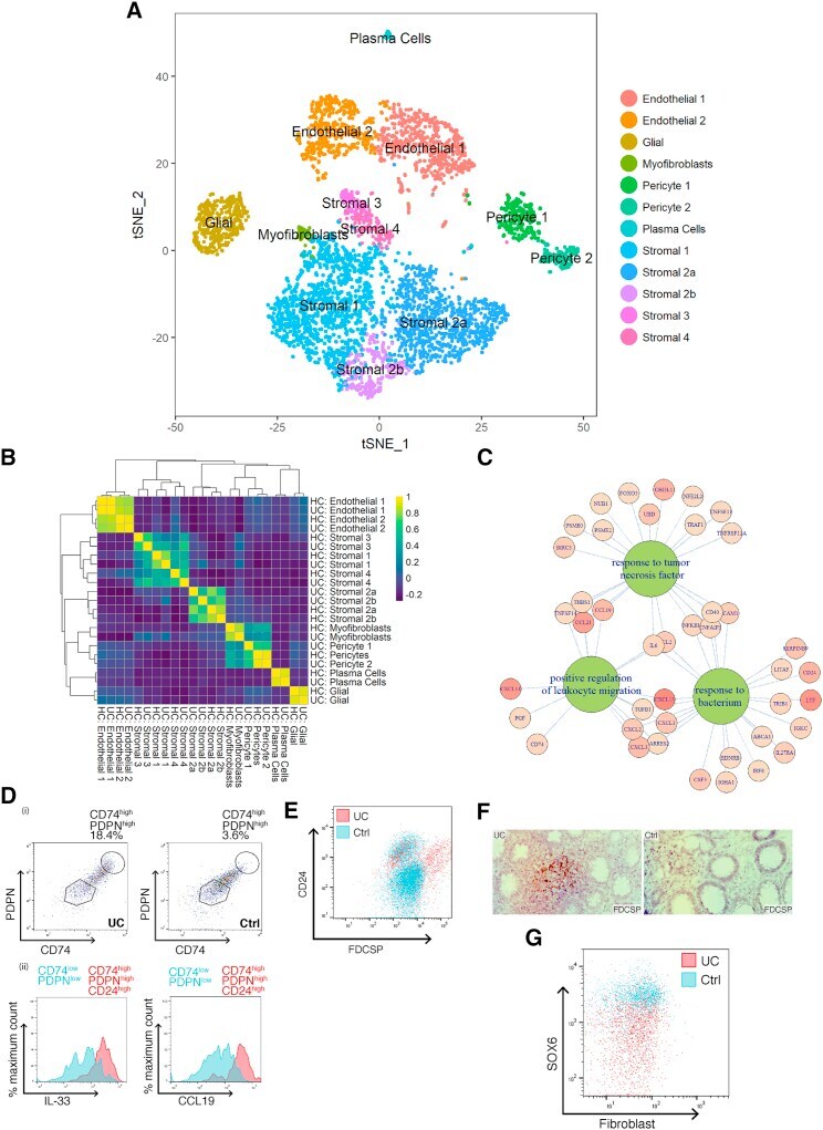
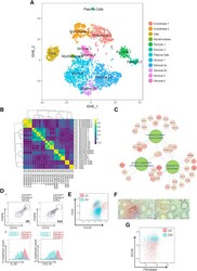
- Figure 2 Colonic Mesenchymal Plasticity in IBD (A) t-SNE plot of UC colonic mesenchyme dataset. Single cells colored by cluster annotation. Descriptive cluster labels are shown. (B) Human healthy and UC cluster marker gene overlap correlation heatmap. (C) Selected enriched (FDR < 0.01) GO terms of UC S4 mesenchymal population marker genes. (D) (i) Flow cytometry analysis of CD74 and PDPN expression on colonic stromal cells from Ctrl (right) or UC (left) donors. (ii) Comparison of intracellular CCL19 and IL-33 levels in CD74 high PDPN high CD24 high cells (red) versus the corresponding CD74 low PDPN low subset (blue) in inflamed UC colonic tissue. (E) Flow cytometry analysis of FDCSP high and CD24 high colonic stromal cells from Ctrl (blue) or UC (red). (F) Single-molecule ISH staining of FDCSP in Ctrl or UC colonic tissue sections. (G) Flow cytometric analysis of SOX6 expression in Ctrl (blue) or UC (red) colonic stromal cells. See also Figures S1 and S3 and Tables S1 and S5 .
- Conjugate
- Green dye