Explore
 Validate
 Learn
Learn Western blot
 Immunocytochemistry
Antibody data
- Antibody Data
- Antigen structure
- References [4]
- Comments [0]
- Validations
- Immunocytochemistry [2]
- Immunohistochemistry [1]
- Other assay [4]
Submit
Validation data
Reference
Comment
Report error
- Product number
- 14-0747-82 - Provider product page

- Provider
- Invitrogen Antibodies
- Product name
- CD74 Monoclonal Antibody (VIC-Y1), eBioscience™
- Antibody type
- Monoclonal
- Antigen
- Other
- Description
- Description: This VIC-Y1 monoclonal antibody recognizes human CD74, also known as the MHC Class II-associated invariant chain. A 33-43 kDa nonpolymorphic type II membrane protein, CD74 is highly expressed on B cells and subsets of activated T cells, Langerhans cells, dendritic cells, and epithelial cells. Monocytes and macrophages also express CD74, but at lower levels. CD74 acts as a chaperone of MHC Class II (HLA-DR) proteins, promoting their translocation from the ER to endocytic compartments during antigen presentation. Activation of NFkB and ERK has been shown following CD74 interaction with CD44 and binding to the proinflammatory cytokine macrophage migration-inhibitory factor (MIF). CD74 also plays a role in the maturation of follicular B-cells and accumulation of marginal zone B-cells. CD74 has also been demonstrated to interact with CXCR2 and CXCR4. The VIC-Y1 monoclonal antibody binds to a cytoplasmic region of human CD74 and thus cannot be used to detect cell surface expression of this molecule. Applications Reported: This VIC-Y1 antibody has been reported for use in immunohistochemical staining of formalin-fixed paraffin embedded tissue sections. Applications Tested: This VIC-Y1 antibody has been tested by immunohistochemistry on formalin-fixed paraffin embedded human tonsil tissue using low pH antigen retrieval. This antibody can be used at less than or equal to 5 µg/mL. It is recommended that the antibody be carefully titrated for optimal performance in the assay of interest. Purity: Greater than 90%, as determined by SDS-PAGE. Aggregation: Less than 10%, as determined by HPLC. Filtration: 0.2 µm post-manufacturing filtered.
- Reactivity
- Human
- Host
- Mouse
- Isotype
- IgG
- Antibody clone number
- VIC-Y1
- Vial size
- 100 μg
- Concentration
- 0.5 mg/mL
- Storage
- 4°C
Submitted references A functional heteromeric MIF receptor formed by CD74 and CXCR4.
CD74 is a member of the regulated intramembrane proteolysis-processed protein family.
Invariant chain targets HLA class II molecules to acidic endosomes containing internalized influenza virus.
Human major histocompatibility complex class II invariant chain is expressed on the cell surface.
Schwartz V, Lue H, Kraemer S, Korbiel J, Krohn R, Ohl K, Bucala R, Weber C, Bernhagen J
FEBS letters 2009 Sep 3;583(17):2749-57
FEBS letters 2009 Sep 3;583(17):2749-57
CD74 is a member of the regulated intramembrane proteolysis-processed protein family.
Becker-Herman S, Arie G, Medvedovsky H, Kerem A, Shachar I
Molecular biology of the cell 2005 Nov;16(11):5061-9
Molecular biology of the cell 2005 Nov;16(11):5061-9
Invariant chain targets HLA class II molecules to acidic endosomes containing internalized influenza virus.
Lamb CA, Yewdell JW, Bennink JR, Cresswell P
Proceedings of the National Academy of Sciences of the United States of America 1991 Jul 15;88(14):5998-6002
Proceedings of the National Academy of Sciences of the United States of America 1991 Jul 15;88(14):5998-6002
Human major histocompatibility complex class II invariant chain is expressed on the cell surface.
Wraight CJ, van Endert P, Möller P, Lipp J, Ling NR, MacLennan IC, Koch N, Moldenhauer G
The Journal of biological chemistry 1990 Apr 5;265(10):5787-92
The Journal of biological chemistry 1990 Apr 5;265(10):5787-92
No comments: Submit comment
Supportive validation
- Submitted by
- Invitrogen Antibodies (provider)
- Main image

- Experimental details
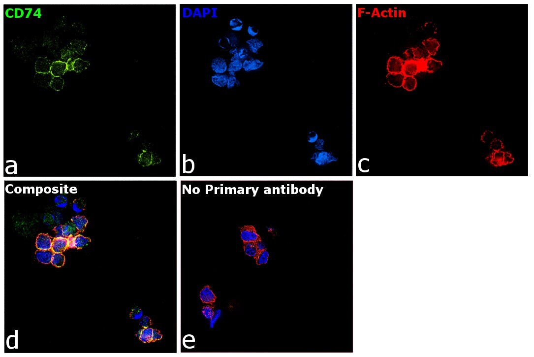
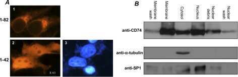
- Immunofluorescence analysis of CD74 was performed using Daudi cells. The cells were fixed with 4% paraformaldehyde for 10 minutes, permeabilized with 0.1% Triton™ X-100 for 15 minutes, and blocked with 2% BSA for 1 hour at room temperature. The cells were labeled with CD74 Monoclonal Antibody (VIC-Y1) (Product # 14-0747-82) at 1:100 dilution in 0.1% BSA, incubated at 4 degree Celsius overnight and then labeled with Donkey anti-Mouse IgG (H+L) Highly Cross-Adsorbed Secondary Antibody, Alexa Fluor Plus 488 (Product # A32766) at a dilution of 1:2000 for 45 minutes at room temperature (Panel a: green).Nuclei (Panel b: blue) were stained with SlowFade® Gold Antifade Mountant with DAPI (Product # S36938). F-actin (Panel c: red) was stained with Rhodamine Phalloidin (Product # R415, 1:300). Panel d represents the merged image showing localization to the plasma membrane. Panel e represents control cells with no primary antibody to assess background. The images were captured at 60X magnification.
- Submitted by
- Invitrogen Antibodies (provider)
- Main image

- Experimental details
- Immunofluorescence analysis of CD74 was performed using Daudi cells. The cells were fixed with 4% paraformaldehyde for 10 minutes, permeabilized with 0.1% Triton™ X-100 for 15 minutes, and blocked with 2% BSA for 1 hour at room temperature. The cells were labeled with CD74 Monoclonal Antibody (VIC-Y1) (Product # 14-0747-82) at 1:100 dilution in 0.1% BSA, incubated at 4 degree Celsius overnight and then labeled with Donkey anti-Mouse IgG (H+L) Highly Cross-Adsorbed Secondary Antibody, Alexa Fluor Plus 488 (Product # A32766) at a dilution of 1:2000 for 45 minutes at room temperature (Panel a: green).Nuclei (Panel b: blue) were stained with SlowFade® Gold Antifade Mountant with DAPI (Product # S36938). F-actin (Panel c: red) was stained with Rhodamine Phalloidin (Product # R415, 1:300). Panel d represents the merged image showing localization to the plasma membrane. Panel e represents control cells with no primary antibody to assess background. The images were captured at 60X magnification.

Supportive validation
- Submitted by
- Invitrogen Antibodies (provider)
- Main image
- Experimental details
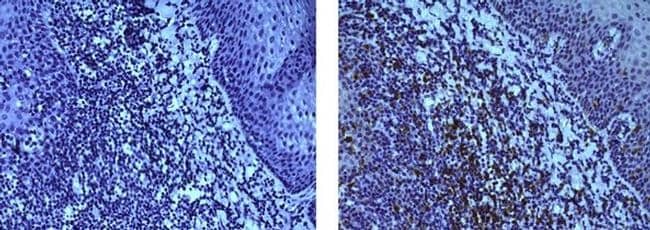
- Immunohistochemistry on formalin-fixed paraffin embedded human tonsil, using 5 µg/mL of Mouse IgG1 kappa Isotype Control (Product # 14-4714-82) (left) or Anti-Human CD74 Purified (right) followed by biotinylated Anti-Mouse IgG Biotin (Product # 13-4013-85) and DAB visualization. Nuclei are counterstained with hematoxylin.
Supportive validation
- Submitted by
- Invitrogen Antibodies (provider)
- Main image
- Experimental details
- NULL
- Submitted by
- Invitrogen Antibodies (provider)
- Main image
- Experimental details
- NULL
- Submitted by
- Invitrogen Antibodies (provider)
- Main image

- Experimental details
- NULL
- Submitted by
- Invitrogen Antibodies (provider)
- Main image
- Experimental details
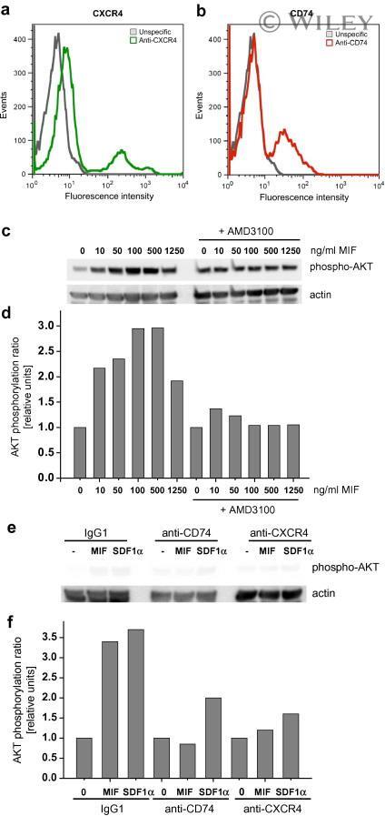
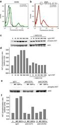
- CXCR4/CD74 receptor complex formation correlates with a functional interplay between CD74 and CXCR4. The CXCR4 inhibitor AMD3100 as well as anti-CXCR4 and anti-CD74 antibodies block MIF-mediated AKT activation in Jurkat T cells, whereas SDF-1alpha-mediated AKT activation is only reduced by anti-CXCR4 but not anti-CD74. (a) Flow cytometry histogram showing surface expression of CXCR4 in Jurkat T cells. Fluorescence intensity of anti-CXCR4-stained cells (green) is compared with that of cells stained with a non-specific FITC-IgG (grey). (b) Flow cytometry histogram showing surface expression of CD74 in Jurkat T cells. Fluorescence intensity of anti-CD74-stained cells (red) is compared with that of cells stained with a non-specific FITC-IgG (grey). (c) Jurkat cells pre-treated with AMD3100 (1 mug/ml, 30 min; +AMD3100) or with control buffer were incubated with rMIF at indicated concentrations for 10 min and AKT activation measured by Western blot using a phospho-Ser473-AKT antibody. Actin staining was performed for standardization of the blot. (d) Quantification of the blot according to (c) (phosphorylation ratio: AKT/actin; mean of n = 2). (e) As in c except that Jurkat cells were pre-treated with anti-CD74, anti-CXCR4 antibody or control IgG1 (10 mug/ml each, 30 min). MIF was added at a concentration of 100 ng/ml and for comparison cells were stimulated with 100 ng/ml SDF-1alpha. (f) Quantification of the blot according to (e) (phosphorylation ratio: AKT/actin; mean of n = 2).