Explore
 Validate
 Learn
Learn Western blot
Antibody data
- Antibody Data
- Antigen structure
- References [24]
- Comments [0]
- Validations
- Western blot [1]
- Immunohistochemistry [1]
- Flow cytometry [2]
- Other assay [7]
Submit
Validation data
Reference
Comment
Report error
- Product number
- MA5-11966 - Provider product page

- Provider
- Invitrogen Antibodies
- Product name
- HLA-DR Monoclonal Antibody (LN3)
- Antibody type
- Monoclonal
- Antigen
- Other
- Description
- MA5-11966 targets Human MHC II (HLA-DR) in FACS, WB and IHC (F) applications and shows reactivity with Human samples. This antibody does not react with mouse tissue in Western blot applications.
- Antibody clone number
- LN3
- Concentration
- 0.2 mg/mL
Submitted references Cuprizone-induced demyelination triggers a CD8-pronounced T cell recruitment.
T cell infiltration in both human multiple system atrophy and a novel mouse model of the disease.
Phase I Study of Ficlatuzumab and Cetuximab in Cetuximab-Resistant, Recurrent/Metastatic Head and Neck Cancer.
Human pDCs Are Superior to cDC2s in Attracting Cytolytic Lymphocytes in Melanoma Patients Receiving DC Vaccination.
Human Inner Ear Immune Activity: A Super-Resolution Immunohistochemistry Study.
Oligodendrocyte degeneration and concomitant microglia activation directs peripheral immune cells into the forebrain.
Tissue Transglutaminase Appears in Monocytes and Macrophages but Not in Lymphocytes in White Matter Multiple Sclerosis Lesions.
Regulation of microglial TMEM119 and P2RY12 immunoreactivity in multiple sclerosis white and grey matter lesions is dependent on their inflammatory environment.
The Human Endolymphatic Sac and Inner Ear Immunity: Macrophage Interaction and Molecular Expression.
The Effect of Chronic Hepatitis B Virus Infection on BDCA3+ Dendritic Cell Frequency and Function.
Effects of B Cell Depletion on Early Mycobacterium tuberculosis Infection in Cynomolgus Macaques.
Particular activation phenotype of T cells expressing HLA-DR but not CD38 in GALT from HIV-controllers is associated with immune regulation and delayed progression to AIDS.
Temporal Dynamics of CD8+ T Cell Effector Responses during Primary HIV Infection.
Trained immunity in newborn infants of HBV-infected mothers.
Evidence for innate immune system activation in HIV type 1-infected elite controllers.
Tissue specificity of decellularized rhesus monkey kidney and lung scaffolds.
Amyloid-β oligomers stimulate microglia through a tyrosine kinase dependent mechanism.
Alzheimer's disease modifies progenitor cell expression of monoamine oxidase B in the subventricular zone.
Dynamics of immune granuloma formation in a Leishmania braziliensis-induced self-limiting cutaneous infection in the primate Macaca mulatta.
Combined therapy of transcatheter hepatic arterial embolization with intratumoral dendritic cell infusion for hepatocellular carcinoma: clinical safety.
Immunohistochemical analysis of orbital connective tissue specimens of patients with active Graves ophthalmopathy.
Phase I study of autologous tumor vaccines transduced with the GM-CSF gene in four patients with stage IV renal cell cancer in Japan: clinical and immunological findings.
Identification of new MHC-restriction elements for presentation of the p210(BCR-ABL) fusion region to human cytotoxic T lymphocytes.
Human macrophage lectin specific for galactose/N-acetylgalactosamine is a marker for cells at an intermediate stage in their differentiation from monocytes into macrophages.
Kaddatz H, Joost S, Nedelcu J, Chrzanowski U, Schmitz C, Gingele S, Gudi V, Stangel M, Zhan J, Santrau E, Greiner T, Frenz J, Müller-Hilke B, Müller M, Amor S, van der Valk P, Kipp M
Glia 2021 Apr;69(4):925-942
Glia 2021 Apr;69(4):925-942
T cell infiltration in both human multiple system atrophy and a novel mouse model of the disease.
Williams GP, Marmion DJ, Schonhoff AM, Jurkuvenaite A, Won WJ, Standaert DG, Kordower JH, Harms AS
Acta neuropathologica 2020 May;139(5):855-874
Acta neuropathologica 2020 May;139(5):855-874
Phase I Study of Ficlatuzumab and Cetuximab in Cetuximab-Resistant, Recurrent/Metastatic Head and Neck Cancer.
Bauman JE, Ohr J, Gooding WE, Ferris RL, Duvvuri U, Kim S, Johnson JT, Soloff AC, Wallweber G, Winslow J, Gaither-Davis A, Grandis JR, Stabile LP
Cancers 2020 Jun 11;12(6)
Cancers 2020 Jun 11;12(6)
Human pDCs Are Superior to cDC2s in Attracting Cytolytic Lymphocytes in Melanoma Patients Receiving DC Vaccination.
van Beek JJP, Flórez-Grau G, Gorris MAJ, Mathan TSM, Schreibelt G, Bol KF, Textor J, de Vries IJM
Cell reports 2020 Jan 28;30(4):1027-1038.e4
Cell reports 2020 Jan 28;30(4):1027-1038.e4
Human Inner Ear Immune Activity: A Super-Resolution Immunohistochemistry Study.
Liu W, Kämpfe Nordström C, Danckwardt-Lillieström N, Rask-Andersen H
Frontiers in neurology 2019;10:728
Frontiers in neurology 2019;10:728
Oligodendrocyte degeneration and concomitant microglia activation directs peripheral immune cells into the forebrain.
Chrzanowski U, Bhattarai S, Scheld M, Clarner T, Fallier-Becker P, Beyer C, Rohr SO, Schmitz C, Hochstrasser T, Schweiger F, Amor S, Horn-Bochtler A, Denecke B, Nyamoya S, Kipp M
Neurochemistry international 2019 Jun;126:139-153
Neurochemistry international 2019 Jun;126:139-153
Tissue Transglutaminase Appears in Monocytes and Macrophages but Not in Lymphocytes in White Matter Multiple Sclerosis Lesions.
Chrobok NL, Bol JGJM, Wilhelmus MMM, Drukarch B, van Dam AM
Journal of neuropathology and experimental neurology 2019 Jun 1;78(6):492-500
Journal of neuropathology and experimental neurology 2019 Jun 1;78(6):492-500
Regulation of microglial TMEM119 and P2RY12 immunoreactivity in multiple sclerosis white and grey matter lesions is dependent on their inflammatory environment.
van Wageningen TA, Vlaar E, Kooij G, Jongenelen CAM, Geurts JJG, van Dam AM
Acta neuropathologica communications 2019 Dec 11;7(1):206
Acta neuropathologica communications 2019 Dec 11;7(1):206
The Human Endolymphatic Sac and Inner Ear Immunity: Macrophage Interaction and Molecular Expression.
Kämpfe Nordström C, Danckwardt-Lillieström N, Laurell G, Liu W, Rask-Andersen H
Frontiers in immunology 2018;9:3181
Frontiers in immunology 2018;9:3181
The Effect of Chronic Hepatitis B Virus Infection on BDCA3+ Dendritic Cell Frequency and Function.
van der Aa E, Buschow SI, Biesta PJ, Janssen HL, Woltman AM
PloS one 2016;11(8):e0161235
PloS one 2016;11(8):e0161235
Effects of B Cell Depletion on Early Mycobacterium tuberculosis Infection in Cynomolgus Macaques.
Phuah J, Wong EA, Gideon HP, Maiello P, Coleman MT, Hendricks MR, Ruden R, Cirrincione LR, Chan J, Lin PL, Flynn JL
Infection and immunity 2016 May;84(5):1301-1311
Infection and immunity 2016 May;84(5):1301-1311
Particular activation phenotype of T cells expressing HLA-DR but not CD38 in GALT from HIV-controllers is associated with immune regulation and delayed progression to AIDS.
Gonzalez SM, Taborda NA, Correa LA, Castro GA, Hernandez JC, Montoya CJ, Rugeles MT
Immunologic research 2016 Jun;64(3):765-74
Immunologic research 2016 Jun;64(3):765-74
Temporal Dynamics of CD8+ T Cell Effector Responses during Primary HIV Infection.
Demers KR, Makedonas G, Buggert M, Eller MA, Ratcliffe SJ, Goonetilleke N, Li CK, Eller LA, Rono K, Maganga L, Nitayaphan S, Kibuuka H, Routy JP, Slifka MK, Haynes BF, McMichael AJ, Bernard NF, Robb ML, Betts MR
PLoS pathogens 2016 Aug;12(8):e1005805
PLoS pathogens 2016 Aug;12(8):e1005805
Trained immunity in newborn infants of HBV-infected mothers.
Hong M, Sandalova E, Low D, Gehring AJ, Fieni S, Amadei B, Urbani S, Chong YS, Guccione E, Bertoletti A
Nature communications 2015 Mar 25;6:6588
Nature communications 2015 Mar 25;6:6588
Evidence for innate immune system activation in HIV type 1-infected elite controllers.
Krishnan S, Wilson EM, Sheikh V, Rupert A, Mendoza D, Yang J, Lempicki R, Migueles SA, Sereti I
The Journal of infectious diseases 2014 Mar;209(6):931-9
The Journal of infectious diseases 2014 Mar;209(6):931-9
Tissue specificity of decellularized rhesus monkey kidney and lung scaffolds.
Nakayama KH, Lee CC, Batchelder CA, Tarantal AF
PloS one 2013;8(5):e64134
PloS one 2013;8(5):e64134
Amyloid-β oligomers stimulate microglia through a tyrosine kinase dependent mechanism.
Dhawan G, Floden AM, Combs CK
Neurobiology of aging 2012 Oct;33(10):2247-61
Neurobiology of aging 2012 Oct;33(10):2247-61
Alzheimer's disease modifies progenitor cell expression of monoamine oxidase B in the subventricular zone.
Pugliese M, Rodríguez MJ, Gimeno-Bayon J, Pujadas L, Billett EE, Wells C, Mahy N
Journal of neuroscience research 2010 Sep;88(12):2588-97
Journal of neuroscience research 2010 Sep;88(12):2588-97
Dynamics of immune granuloma formation in a Leishmania braziliensis-induced self-limiting cutaneous infection in the primate Macaca mulatta.
Souza-Lemos C, de-Campos SN, Teva A, Côrte-Real S, Fonseca EC, Porrozzi R, Grimaldi G Jr
The Journal of pathology 2008 Nov;216(3):375-86
The Journal of pathology 2008 Nov;216(3):375-86
Combined therapy of transcatheter hepatic arterial embolization with intratumoral dendritic cell infusion for hepatocellular carcinoma: clinical safety.
Nakamoto Y, Mizukoshi E, Tsuji H, Sakai Y, Kitahara M, Arai K, Yamashita T, Yokoyama K, Mukaida N, Matsushima K, Matsui O, Kaneko S
Clinical and experimental immunology 2007 Feb;147(2):296-305
Clinical and experimental immunology 2007 Feb;147(2):296-305
Immunohistochemical analysis of orbital connective tissue specimens of patients with active Graves ophthalmopathy.
Avunduk AM, Avunduk MC, Pazarli H, Oguz V, Varnell ED, Kaufman HE, Aksoy F
Current eye research 2005 Aug;30(8):631-8
Current eye research 2005 Aug;30(8):631-8
Phase I study of autologous tumor vaccines transduced with the GM-CSF gene in four patients with stage IV renal cell cancer in Japan: clinical and immunological findings.
Tani K, Azuma M, Nakazaki Y, Oyaizu N, Hase H, Ohata J, Takahashi K, OiwaMonna M, Hanazawa K, Wakumoto Y, Kawai K, Noguchi M, Soda Y, Kunisaki R, Watari K, Takahashi S, Machida U, Satoh N, Tojo A, Maekawa T, Eriguchi M, Tomikawa S, Tahara H, Inoue Y, Yoshikawa H, Yamada Y, Iwamoto A, Hamada H, Yamashita N, Okumura K, Kakizoe T, Akaza H, Fujime M, Clift S, Ando D, Mulligan R, Asano S
Molecular therapy : the journal of the American Society of Gene Therapy 2004 Oct;10(4):799-816
Molecular therapy : the journal of the American Society of Gene Therapy 2004 Oct;10(4):799-816
Identification of new MHC-restriction elements for presentation of the p210(BCR-ABL) fusion region to human cytotoxic T lymphocytes.
Sun JY, Senitzer D, Forman SJ, Chatterjee S, Wong KK Jr
Cancer immunology, immunotherapy : CII 2003 Dec;52(12):761-70
Cancer immunology, immunotherapy : CII 2003 Dec;52(12):761-70
Human macrophage lectin specific for galactose/N-acetylgalactosamine is a marker for cells at an intermediate stage in their differentiation from monocytes into macrophages.
Higashi N, Morikawa A, Fujioka K, Fujita Y, Sano Y, Miyata-Takeuchi M, Suzuki N, Irimura T
International immunology 2002 Jun;14(6):545-54
International immunology 2002 Jun;14(6):545-54
No comments: Submit comment
Supportive validation
- Submitted by
- Invitrogen Antibodies (provider)
- Main image

- Experimental details
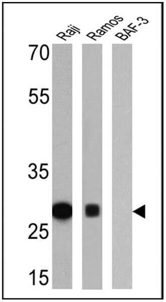
- Western blot analysis of Human MHC II (HLA-DR) was performed by loading 25 µg of Raji (lane 1), Ramos (lane 2) and BAF-3 (lane 3) cell lysates onto an SDS polyacrylamide gel. Proteins were transferred to a PVDF membrane and blocked at 4ºC overnight. The membrane was probed with a Human MHC II (HLA-DR) monoclonal antibody (Product # MA5-11966) at a dilution of 1:10,000 (Raji) and 1:200 (Ramos and BAF-3) overnight at 4°C, washed in TBST, and probed with an HRP-conjugated secondary antibody for 1 hr at room temperature in the dark. Chemiluminescent detection was performed using Pierce ECL Plus Western Blotting Substrate (Product # 32132). Results show a band at ~27 kDa in Raji and Ramos cells.
Supportive validation
- Submitted by
- Invitrogen Antibodies (provider)
- Main image
- Experimental details
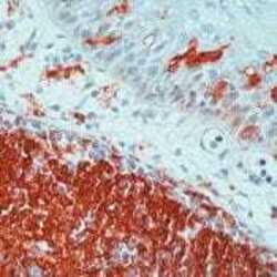
- Formalin-fixed, paraffin-embedded human tonsil stained with HLA-DR la antibody using peroxidase-conjugate and AEC chromogen. Note membrane staining of B cells and macrophages

Supportive validation
- Submitted by
- Invitrogen Antibodies (provider)
- Main image
- Experimental details
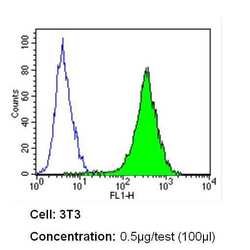
- Flow cytometry analysis of Human MHC II (HLA-DR) in NIH-3T3 cells (green) compared to an isotype control (blue). Cells were harvested, adjusted to a concentration of 1-5x10^6 cells/mL, fixed with 2% paraformaldehyde and washed with PBS. Cells were blocked with a 2% solution of BSA-PBS for 30 min at room temperature and incubated with a Human MHC II (HLA-DR) monoclonal antibody (Product # MA5-11966) at a dilution of 0.5 µg/test for 60 min at room temperature. Cells were then incubated for 40 min at room temperature in the dark using a Dylight 488-conjugated secondary antibody and re-suspended in PBS for FACS analysis.

- Submitted by
- Invitrogen Antibodies (provider)
- Main image

- Experimental details
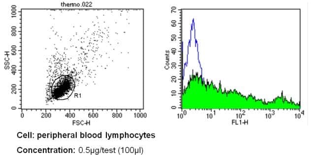
- Flow cytometry analysis of Human MHC II (HLA-DR) in PBMC cells (green) compared to an isotype control (blue). Human blood was collected, combined with a hydrophilic polysaccharide, centrifuged, transferred to a conical tube and washed with PBS. 50 µL of cell solution was added to each tube at a dilution of 2x10^7 cells/mL, followed by the addition of 50 µL of isotype control and primary antibody (Product # MA5-11966) at a dilution of 0.5 µg/test. Cells were incubated for 30 min at 4ºC and washed with a cell buffer, followed by incubation with a DyLight 488-conjugated secondary antibody for 30 min at 4ºC in the dark. FACS analysis was performed using 400 µL of cell buffer.
Supportive validation
- Submitted by
- Invitrogen Antibodies (provider)
- Main image
- Experimental details
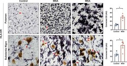
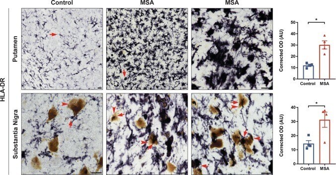
- Fig. 1 HLA-DR expression in human postmortem MSA brain tissue. Control (left) and MSA (middle and right) postmortem brain sections of the putamen (top) and substantia nigra (bottom) stained with HLA-DR (DAB, black staining). Quantification of the staining intensity revealed a significant increase of HLA-DR staining in the putamen and in the substantia nigra. Arrows denote ""resting"" (control, left) or ""activated"" (MSA, middle and right) microglia; arrowheads denote neuromelanin laden dopamine neurons in the substantia nigra. Representative images. All scale bars indicate 25 mum. Graphs display the mean +- SEM. * p < 0.05, unpaired nonparametric Mann Whitney Test was used to compare groups, n = 4 per group
- Submitted by
- Invitrogen Antibodies (provider)
- Main image
- Experimental details
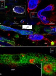
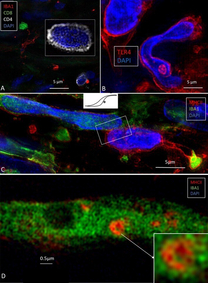
- Figure 2 (A) SR-SIM of CD4- and CD8-positive cells present in the perisaccular tissue. (B) Some cells express the toll-like receptor 4 (TLR4). (C) Sub-epithelial cell interaction near the external aperture of the vestibular aqueduct. IBA1 cells interact (framed area) with cells strongly expressing MHCII. Cell nuclei show different protein expression [from Kampfe-Nordstrom et al. () with permission]. (D) A sub-epithelial IBA1 cell contains a multi-vesicular body expressing MHCII.
- Submitted by
- Invitrogen Antibodies (provider)
- Main image
- Experimental details
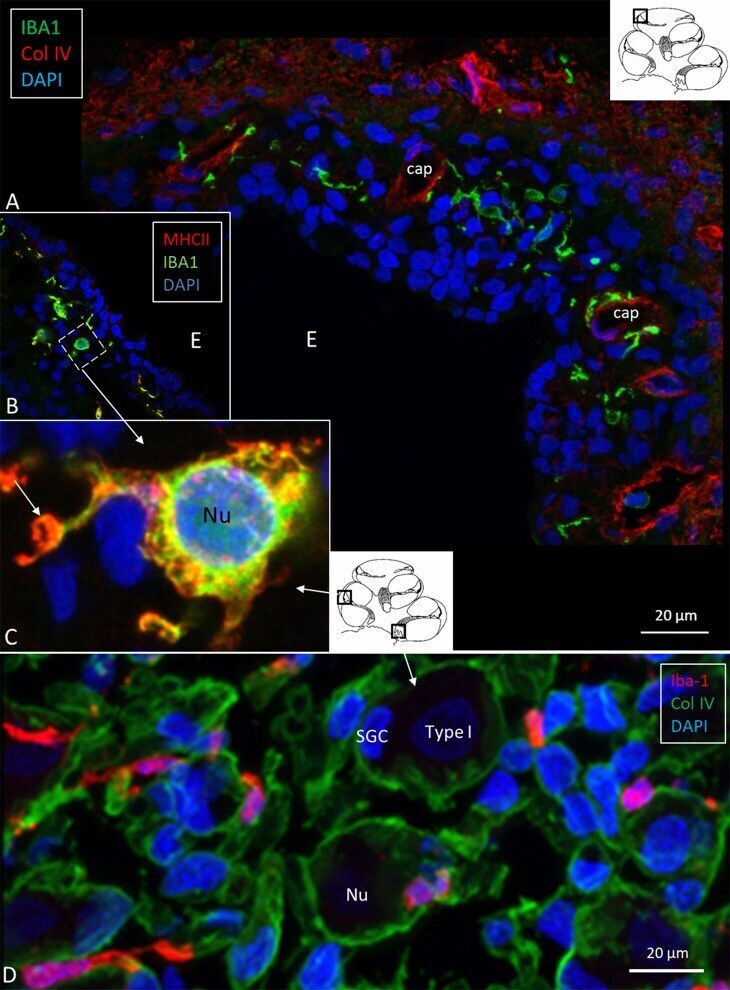
- Figure 3 (A) Immunofluorescence of IBA1 and collagen IV in the lateral wall of the apical turn of the human cochlea. Many perivascular IBA1 cells are seen in the StV and few in the spiral ligament. (B) Confocal microscopy of the human StV. Framed area is magnified in (C) . (C) SR-SIM of framed area in B. Cell co-express IBA1 and MHCII. The cell membrane expresses MHCII as well as cytoplasmic vesicles [ (B,C) from Kampfe-Nordstrom et al. ( 12 )]. (D) Confocal microscopy of spiral ganglion with several surrounding IBA1 cells. SGC, satellite glial cell; Nu, type I cell nucleus; Col. IV, collagen IV; cap, capillary; E, endolymph.
- Submitted by
- Invitrogen Antibodies (provider)
- Main image
- Experimental details
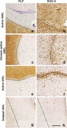
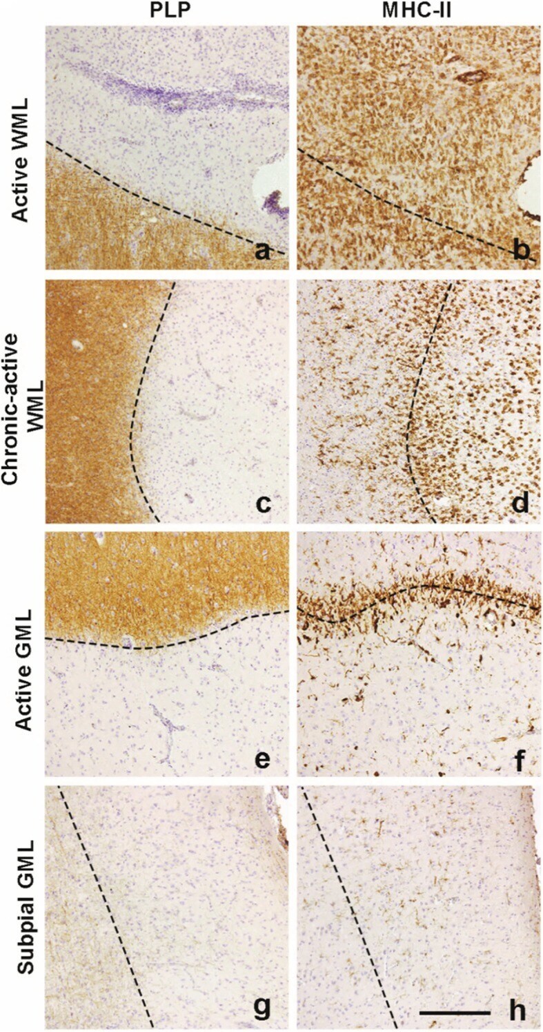
- Fig. 1 Representative images of lesion types used in this study. Lesions are characterized by loss of PLP staining and amount of MHC-II+ cells. A large amount of of MHC-II+ cells can be observed in the demyelinated area ( a ) in active WMLs ( b ). Chronic-active demyelinated WMLs ( c ) feature a 'rim' of MHC-II cells ( d ) which is also visible in demyelinated active GMLs ( e, f ). Subpial demyelinated ( g ) GMLs hardly show MHC-II+ cells ( h ). Scalebar ( a-h ) = 200 mum. Dashed lines indicate the edge of the lesion
- Submitted by
- Invitrogen Antibodies (provider)
- Main image
- Experimental details
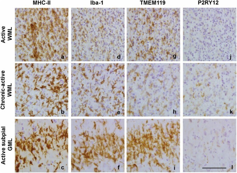
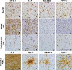
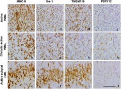
- Fig. 2 Representative images of MHC-II ( a, b, c ), Iba-1 ( d, e, f ), TMEM119 ( g, h, i ) and P2RY12 ( j, k, l ) immunoreactivity in normal appearing matter and in the demyelinated center of active WMLs and chronic-active WMLs and in pre-active lesions ( m, n, o, p ). Scalebars ( a-l and m-p ) = 50 mum

- Submitted by
- Invitrogen Antibodies (provider)
- Main image
- Experimental details
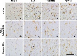
- Fig. 3 Representative images of MHC-II ( a, b, c ), Iba-1 ( d, e, f ), TMEM119 ( g, h, i ) and P2RY12 ( i, k,l ) immunoreactivity in NAGM, active subpial GMLs and non-active subpial GMLs. Arrows indicate rod-shaped microglia visible in subpial GMLs in Iba-1+ cells ( f ), TMEM119 + cells ( i ) and P2RY12+ cells ( l ). Scalebar = 50 mum
- Submitted by
- Invitrogen Antibodies (provider)
- Main image
- Experimental details
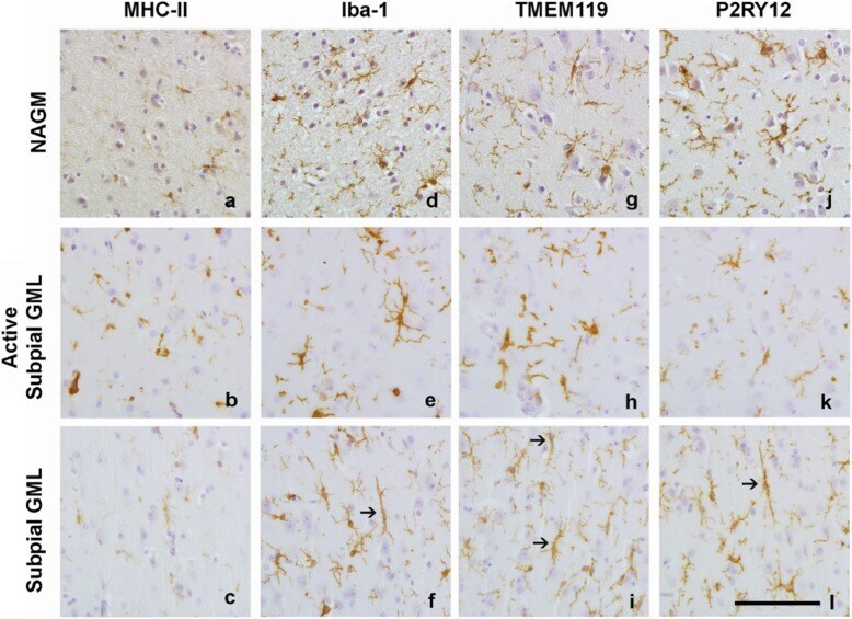
- Fig. 5 Representative images of immunoreactivity for MHC-II ( a, b, c ), Iba-1 ( d, e, f ), TMEM119 ( g, h, i ) and P2RY11 ( j, k, l ) along the rim of various lesion types. Scalebar ( a-l ) = 50 mum