Explore
 Validate
 Learn
Learn Immunohistochemistry
Antibody data
- Antibody Data
- Antigen structure
- References [0]
- Comments [0]
- Validations
- Immunohistochemistry [2]
Submit
Validation data
Reference
Comment
Report error
- Product number
- 752-9956-94 - Provider product page

- Provider
- Invitrogen Antibodies
- Product name
- HLA-DR Monoclonal Antibody (LN3), Alexa Fluor™ Plus 488
- Antibody type
- Monoclonal
- Antigen
- Other
- Description
- Description: The LN3 antibody reacts with the human major histocompatibility complex (MHC) class II, HLA-DR. HLA-DR is expressed on the surface of human antigen presenting cells (APC) including B cells, monocytes, macrophages, DCs, and activated T cells. HLA-DR is a heterodimeric transmembrane protein composed of alpha and beta subunits and plays an important role in the presentation of peptides to CD4^+ T lymphocytes. Applications Reported: The LN3 antibody has been reported for use in flow cytometric analysis, and immunohistochemical staining. Immunohistochemistry can be performed using frozen and Bouin's, or formalin/paraffin, human tissues. Applications Tested: This LN3 antibody has been tested by immunohistochemistry of formalin-fixed paraffin embedded tissue using high pH antigen retrieval and can be used at 10 µg/mL. It is recommended that the antibody be carefully titrated for optimal performance in the assay of interest. Using conjugate solutions: Centrifuge the protein conjugate solution briefly in a microcentrifuge before use; add only the supernatant to the experiment. This step will help eliminate any protein aggregates that may have formed during storage, thereby reducing nonspecific background staining.
- Reactivity
- Human
- Host
- Mouse
- Conjugate
- Green dye
- Isotype
- IgG
- Antibody clone number
- LN3
- Vial size
- 500 µL
- Concentration
- 0.2 mg/mL
- Storage
- 4°C, store in dark
No comments: Submit comment
Supportive validation
- Submitted by
- Invitrogen Antibodies (provider)
- Main image
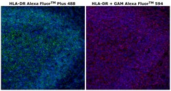
- Experimental details
- Immunohistochemical analysis of HLA-DR was performed on FFPE human tonsil tissue. To expose the target protein, HIER was performed on de-paraffinized sections using BOND Epitope Retrieval Solution 2 (pH 9) for 10 min, followed by a 5 min cool down, a 1 min wash with ddH2O, and then transferred to 1X PBS. Tissues were permeabilized with 0.1% Triton X-100 in 1X PBS for 30 min, washed three times in 1X PBS and blocked with 3% BSA/5% normal goat serum/1X PBS for 1 hr at RT. Tissues were then probed with HLA-DR Monoclonal Antibody (LN3), Alexa Fluor™ Plus 488 (Product # 752-9956-94) at 10 µg/mL (left) or with HLA-DR Monoclonal Antibody (LN3) (Product # MA5-11966) at 1 µg/mL (right), respectively, in blocking solution for 1 hr at RT in a humidified chamber. Tissues were washed twice for 5 min each in 1X PBST (0.05% Tween-20), then once for 5 min in 1X PBS. Detection of the unconjugated primary antibody was performed using Goat anti-Mouse IgG (H+L) Secondary Antibody, Alexa Fluor™ Plus 594 (Product # A32742) at a dilution of 1:2,000 in blocking solution for 1 hr at RT, followed by two 5 min washes in 1X PBST then one 5 min wash in 1X PBS, and then stained with DAPI (Product # 62247) at 1 µg/mL diluted in 1X PBS for 5 min at RT. The sections were washed twice in 1X PBS followed by a final rinse in ddH2O and were mounted with ProLong™ Glass Antifade Mountant (Product # P36982). Images were captured on EVOS™ M7000 Imaging System (Product # AMF7000) at 20X magnification.

- Submitted by
- Invitrogen Antibodies (provider)
- Main image
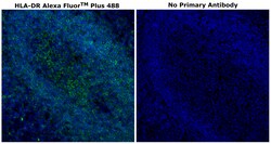
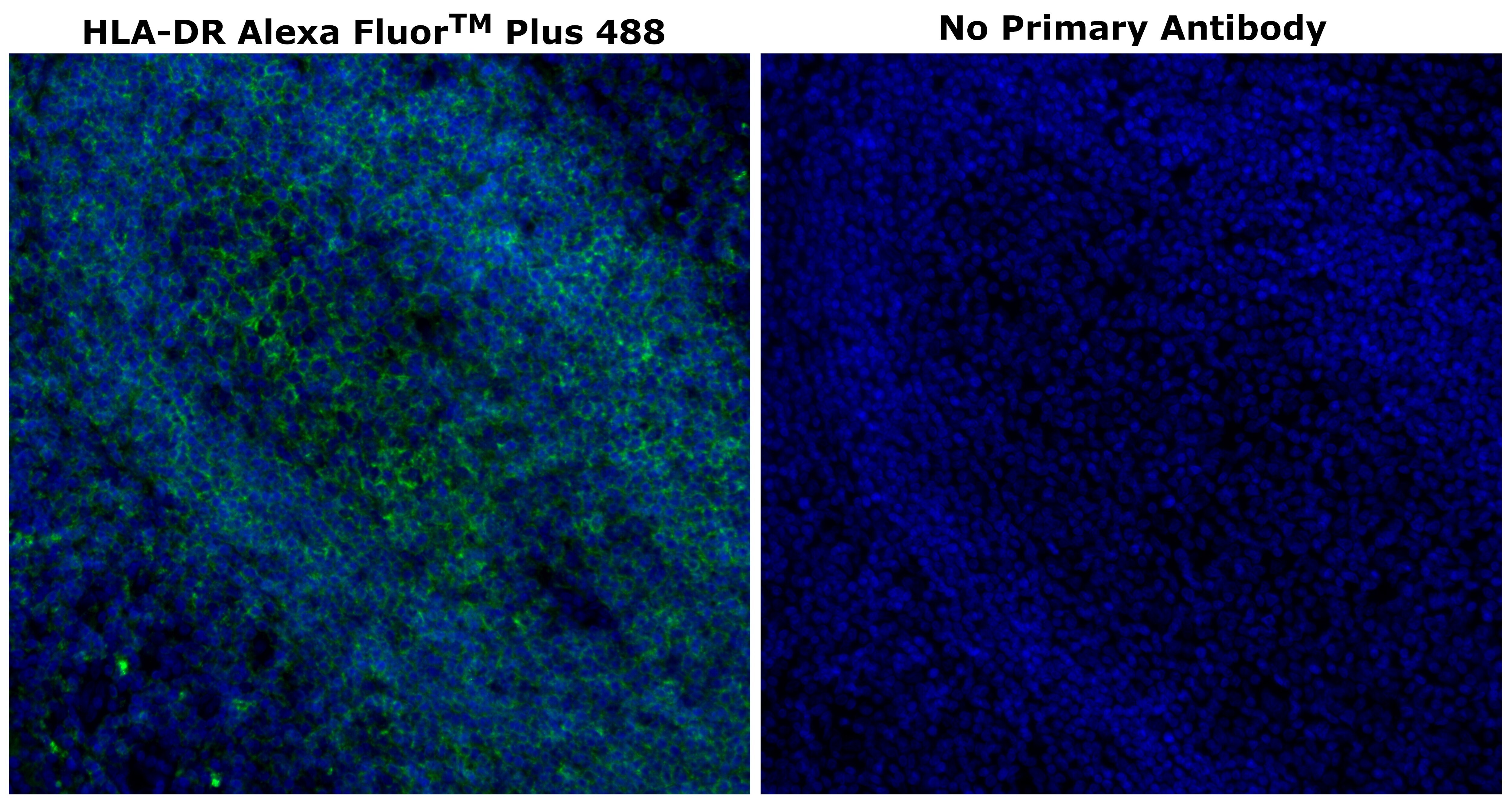
- Experimental details
- Immunohistochemical analysis of HLA-DR was performed on formalin-fixed paraffin-embedded human tonsil tissue. To expose the target protein, HIER was performed on de-paraffinized sections using BOND Epitope Retrieval Solution 2 (pH 9) for 10 min, followed by a 5 min cool down, a 1 min wash with ddH2O, and then transferred to 1X PBS. Tissues were permeabilized with 0.1% Triton X-100 in 1X PBS for 30 min, washed three times in 1X PBS and blocked with 3% BSA/5% normal goat serum/1X PBS for 1 hr at RT. Following the removal of the blocking solution, tissues were probed with (left) or without (right) HLA-DR Monoclonal Antibody (LN3), Alexa Fluor™ Plus 488 (Product # 752-9956-94) at 10 µg/mL in blocking solution for 1 hr at RT in a humidified chamber. Tissues were then washed twice for 5 min each, in 1X PBS + 0.05% Tween-20 (PBST), and once for 5 min in 1X PBS. Tissues were then stained with DAPI (Product # 62247) at 1 µg/mL diluted in 1X PBS for 5 min at RT. The sections were washed twice in 1X PBS followed by a final rinse in ddH2O and were mounted with ProLong™ Glass Antifade Mountant (Product # P36982). Images were captured on EVOS™ M7000 Imaging System (Product # AMF7000) at 20X magnification.