Explore
 Validate
 Learn
Learn Western blot
 Immunocytochemistry
Antibody data
- Antibody Data
- Antigen structure
- References [0]
- Comments [0]
- Validations
- Immunocytochemistry [1]
- Immunoprecipitation [1]
- Other assay [1]
Submit
Validation data
Reference
Comment
Report error
- Product number
- PA5-53581 - Provider product page

- Provider
- Invitrogen Antibodies
- Product name
- Spastin Polyclonal Antibody
- Antibody type
- Polyclonal
- Antigen
- Recombinant protein fragment
- Description
- Immunogen sequence: KRKDPLTHTS NSLPRSKTVM KTGSAGLSGH HRAPSYSGLS MVSGVKQGSG PAPTTHKGTP KTNRTNKPST PTTATRKKKD LKNFRNVDSN LANLIMNEIV DNGTAVKFDD IAGQDLAKQA LQEIVILPSL RPELFTGL Highest antigen sequence identity to the following orthologs: Mouse - 91%, Rat - 91%.
- Reactivity
- Human
- Host
- Rabbit
- Isotype
- IgG
- Vial size
- 100 μL
- Concentration
- 0.20 mg/mL
- Storage
- Store at 4°C short term. For long term storage, store at -20°C, avoiding freeze/thaw cycles.
No comments: Submit comment
Supportive validation
- Submitted by
- Invitrogen Antibodies (provider)
- Main image
- Experimental details
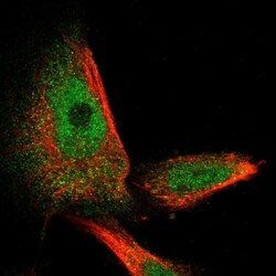
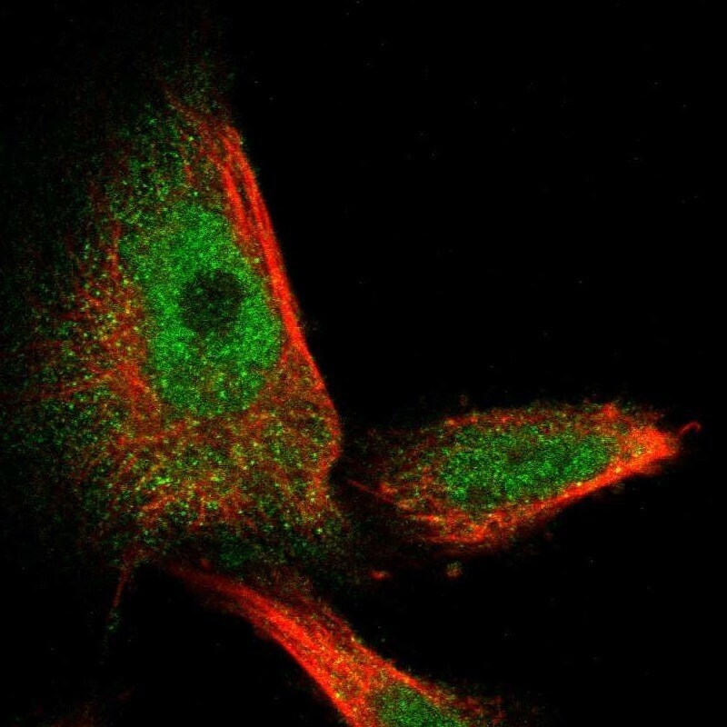
- Immunofluorescent staining of Spastin in human cell line U-251 MG using a Spastin Polyclonal Antibody (Product # PA5-53581) shows localization to nucleoplasm and cytosol.
Supportive validation
- Submitted by
- Invitrogen Antibodies (provider)
- Main image

- Experimental details
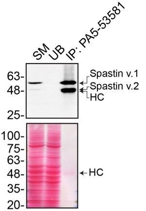
- Immunoprecipitation of SPAST was performed on HAP1 cell lysates. Antibody-bead conjugates were prepared by adding 1 µg of SPAST polyclonal antibody (Product # PA5-53581) with 30 µL of protein A-Sepharose beads and rocked overnight at 4°C. 1 mg of lysate was incubated with an antibody-bead conjugate for 2 hours at 4°C. Following centrifugation and multiple washes, 10% starting material (SM), 10% unbound fraction (UB) and immunoprecipitated fraction (IP) were processed for immunoblot using a different SPAST monoclonal antibody. Ponceau stained transfer of blot is shown. Data courtesy of YCharOS Inc., an open science company with the mission of characterizing commercially available antibodies using knockout validation.

Supportive validation
- Submitted by
- Invitrogen Antibodies (provider)
- Main image

- Experimental details
- Immunoprecipitation of SPAST was performed on HAP1 cell lysates. Antibody-bead conjugates were prepared by adding 1 µg of SPAST polyclonal antibody (Product # PA5-53581) with 30 µL of protein A-Sepharose beads and rocked overnight at 4°C. 1 mg of lysate was incubated with an antibody-bead conjugate for 2 hours at 4°C. Following centrifugation and multiple washes, 10% starting material (SM), 10% unbound fraction (UB) and immunoprecipitated fraction (IP) were processed for immunoblot using a different SPAST monoclonal antibody. Ponceau stained transfer of blot is shown. Data courtesy of YCharOS Inc., an open science company with the mission of characterizing commercially available antibodies using knockout validation.