Explore
 Validate
 Learn
Learn Western blot
Antibody data
- Antibody Data
- Antigen structure
- References [0]
- Comments [0]
- Validations
- Western blot [3]
- Immunohistochemistry [1]
Submit
Validation data
Reference
Comment
Report error
- Product number
- ABIN1504201 - Provider product page

- Provider
- antibodies-online
- Product name
- anti-V-Ha-Ras Harvey Rat Sarcoma Viral Oncogene Homolog (HRAS) antibody
- Antibody type
- Monoclonal
- Antigen
- Other
- Description
- Protein G Chromatography
- Reactivity
- Human, Mouse, Rat
- Host
- Mouse
- Isotype
- IgG
- Antibody clone number
- '1A6-2
- Vial size
- 0.1 mg
- Concentration
- 1.0 mg/mL
No comments: Submit comment
Supportive validation
- Submitted by
- antibodies-online (provider)
- Main image

- Experimental details
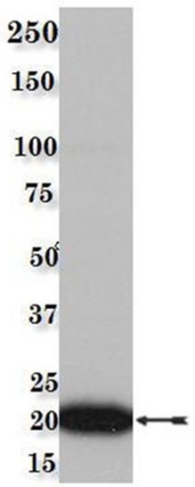
- Western Blot Analysis:Rat brain lysate was resolved by SDS-PAGE, transferred to PVDF, and probed with anti-Ras, clone 1A6.2 (1/1,000). Proteins were visualized using a goat anti-mouse secondary antibody conjugated to HRP and a chemiluminescence detection system. Arrow indicates Ras.
- Submitted by
- antibodies-online (provider)
- Main image

- Experimental details
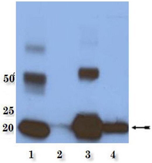
- Ras Isoform Western Blot Analysis:Representative lot data. Recombinant N-Ras, K-Ras, and H-Ras proteins and A431 lysate (Lanes 1, 2, 3, and 4, respectively) were resolved by SDS-PAGE, transferred to PVDF, and probed with anti-Ras, clone 1A6. Proteins were visualized using a goat anti-mouse secondary antibody conjugated to HRP and a chemiluminescence detection system. Arrows indicates Ras.
- Submitted by
- antibodies-online (provider)
- Main image

- Experimental details
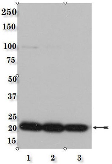
- Species Cross-Reactivity Western Blot Analysis:Mouse brain (lane 1), Rat Brain (lane 2), and A431 (lane 3) cell lysates were resolved by SDS-PAGE, transferred to PVDF, and probed with anti-Ras, clone 1A6. Proteins were visualized using a goat anti-mouse secondary antibody conjugated to HRP and a chemiluminescence detection system. Arrow indicates Ras.
Supportive validation
- Submitted by
- antibodies-online (provider)
- Main image

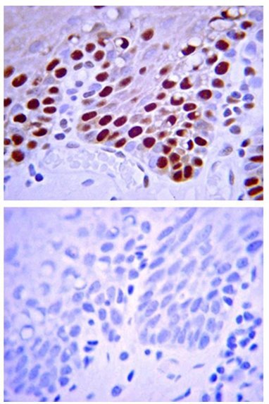
- Experimental details
- Immunohistochemistry:Top: Representative lot data. Representative staining pattern/morphology on stratified Squamous epithelium in human tonsil. Tissue was pretreated with Citrate pH 6.0, antigen retrieval. Antibody was diluted to 1:50, IHC Select Detection System HRP-DAB.Bottom:Representative lot data. Negative staining control representative staining pattern/morphology on stratified Squamous epithelium in human tonsil. Tissue pretreated with Citrate pH 6.0, antigen retrieval. Antibody was omitted, IHC-Select Detection with HRPDAB. Immunoreactivity was not detectable.