Explore
 Validate
 Learn
Learn Western blot
 Immunocytochemistry
Antibody data
- Antibody Data
- Antigen structure
- References [1]
- Comments [0]
- Validations
- Western blot [1]
- Immunohistochemistry [12]
Submit
Validation data
Reference
Comment
Report error
- Product number
- NBP1-86893 - Provider product page

- Provider
- Novus Biologicals
- Proper citation
- Novus Cat#NBP1-86893, RRID:AB_11025576
- Product name
- Rabbit Polyclonal TRP2 Antibody
- Antibody type
- Polyclonal
- Description
- Immunogen affinity purified. Specificity of human TRP2 antibody verified on a Protein Array containing target protein plus 383 other non-specific proteins.
- Reactivity
- Human
- Host
- Rabbit
- Isotype
- IgG
- Vial size
- 0.1 ml
- Storage
- Store at 4C short term. Aliquot and store at -20C long term. Avoid freeze-thaw cycles.
Submitted references Absence of recognition of common melanocytic antigens by T cells isolated from the cerebrospinal fluid of a Vogt-Koyanagi-Harada patient.
Abad S, Wieërs G, Colau D, Wildmann C, Delair E, Dhote R, Brézin AP, Kawakami Y, Coulie PG, van der Bruggen P
Molecular vision 2014;20:956-69
Molecular vision 2014;20:956-69
No comments: Submit comment
Supportive validation
- Submitted by
- Novus Biologicals (provider)
- Main image
- Experimental details
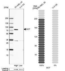
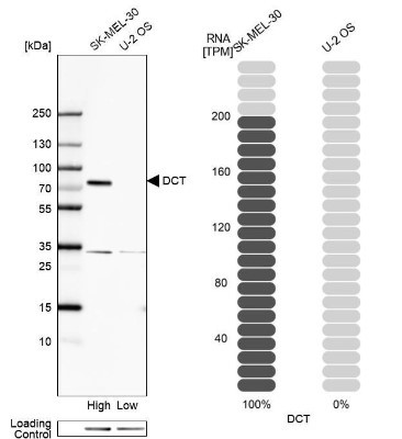
- Western Blot: TRP2 Antibody [NBP1-86893] - Analysis in human cell line SK-MEL-30 and human cell line U-2 OS.
Supportive validation
- Submitted by
- Novus Biologicals (provider)
- Main image
- Experimental details
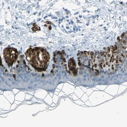
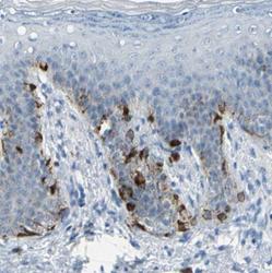
- Immunohistochemistry-Paraffin: TRP2 Antibody [NBP1-86893] - Staining of human skin shows strong cytoplasmic positivity in melanocytes.

- Submitted by
- Novus Biologicals (provider)
- Main image
- Experimental details
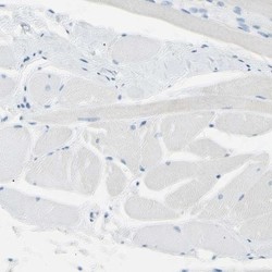
- Immunohistochemistry-Paraffin: TRP2 Antibody [NBP1-86893] - Staining of human skeletal muscle shows low expression as expected.

- Submitted by
- Novus Biologicals (provider)
- Main image
- Experimental details
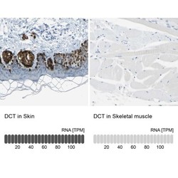
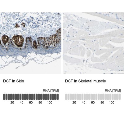
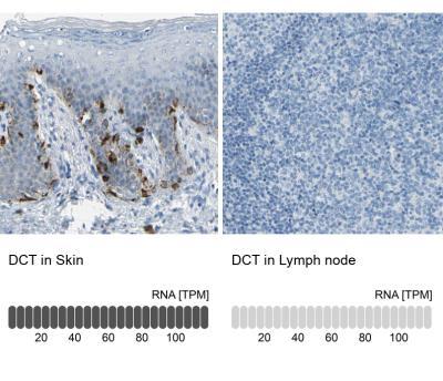
- Immunohistochemistry-Paraffin: TRP2 Antibody [NBP1-86893] - Staining in human skin and skeletal muscle tissues using anti-DCT antibody. Corresponding DCT RNA-seq data are presented for the same tissues.
- Submitted by
- Novus Biologicals (provider)
- Main image
- Experimental details
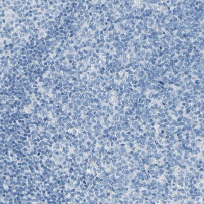
- Immunohistochemistry-Paraffin: TRP2 Antibody [NBP1-86893] - Staining of human lymph node.
- Submitted by
- Novus Biologicals (provider)
- Main image

- Experimental details
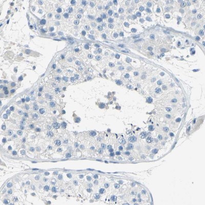
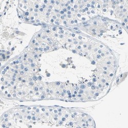
- Immunohistochemistry-Paraffin: TRP2 Antibody [NBP1-86893] - Staining of human testis.
- Submitted by
- Novus Biologicals (provider)
- Main image
- Experimental details
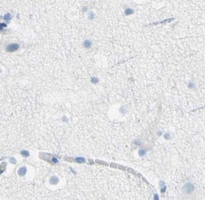
- Immunohistochemistry-Paraffin: TRP2 Antibody [NBP1-86893] - Staining of human cerebral cortex using Anti-DCT antibody.
- Submitted by
- Novus Biologicals (provider)
- Main image
- Experimental details
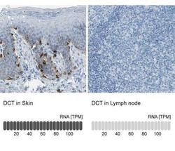
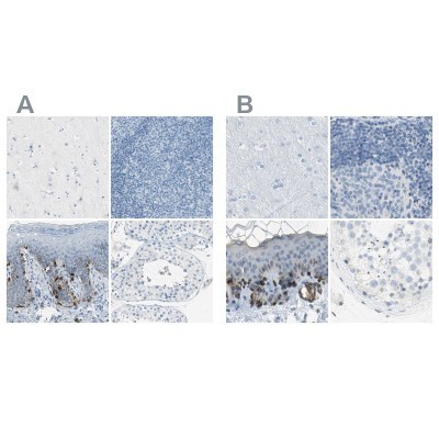
- Immunohistochemistry-Paraffin: TRP2 Antibody [NBP1-86893] - Analysis in human skin and lymph node tissues. Corresponding DCT RNA-seq data are presented for the same tissues.
- Submitted by
- Novus Biologicals (provider)
- Main image
- Experimental details
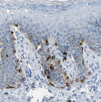
- Immunohistochemistry-Paraffin: TRP2 Antibody [NBP1-86893] - Staining of human skin shows cytoplasmic positivity in squamous epithelial cells.
- Submitted by
- Novus Biologicals (provider)
- Main image
- Experimental details
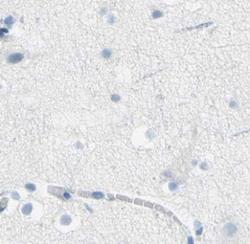
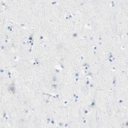
- Immunohistochemistry-Paraffin: TRP2 Antibody [NBP1-86893] - Staining of human cerebral cortex shows no positivity in neurons as expected.

- Submitted by
- Novus Biologicals (provider)
- Main image
- Experimental details
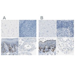
- Immunohistochemistry-Paraffin: TRP2 Antibody [NBP1-86893] - Staining of human cerebral cortex, lymph node, skin and testis using Anti-TRP2 antibody NBP1-86893 (A) shows similar protein distribution across tissues to independent antibody NBP1-86892 (B).
- Submitted by
- Novus Biologicals (provider)
- Main image
- Experimental details
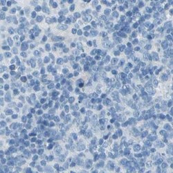
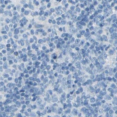
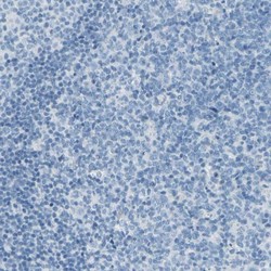
- Immunohistochemistry-Paraffin: TRP2 Antibody [NBP1-86893] - Staining of human lymph node shows no positivity in germinal center cells as expected.
- Submitted by
- Novus Biologicals (provider)
- Main image
- Experimental details
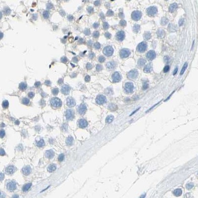
- Immunohistochemistry-Paraffin: TRP2 Antibody [NBP1-86893] - Staining of human testis shows no positivity in cells in seminiferous ducts as expected.