Explore
 Validate
 Learn
Learn Western blot
 Immunohistochemistry
Antibody data
- Antibody Data
- Antigen structure
- References [1]
- Comments [0]
- Validations
- Immunohistochemistry [1]
Submit
Validation data
Reference
Comment
Report error
- Product number
- HPA010743 - Provider product page

- Provider
- Atlas Antibodies
- Proper citation
- Atlas Antibodies Cat#HPA010743, RRID:AB_1847519
- Product name
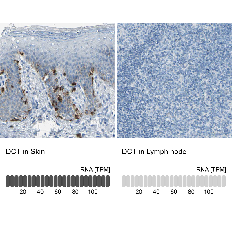
- Anti-DCT
- Antibody type
- Polyclonal
- Description
- Polyclonal Antibody against Human DCT, Gene description: dopachrome tautomerase, Alternative Gene Names: TYRP2, Validated applications: WB, IHC, Uniprot ID: P40126, Storage: Store at +4°C for short term storage. Long time storage is recommended at -20°C.
- Reactivity
- Human
- Host
- Rabbit
- Conjugate
- Unconjugated
- Isotype
- IgG
- Vial size
- 100 µl
- Concentration
- 0.2 mg/ml
- Storage
- Store at +4°C for short term storage. Long time storage is recommended at -20°C.
- Handling
- The antibody solution should be gently mixed before use.
Submitted references Mitochondrial signatures shape phenotype switching and apoptosis in response to PLK1 inhibitors
Lavallée É, Roulet-Matton M, Giang V, Cardona Hurtado R, Chaput D, Gravel S
Life Science Alliance 2025;8(3):e202402912
Life Science Alliance 2025;8(3):e202402912
No comments: Submit comment
Supportive validation
- Submitted by
- Atlas Antibodies (provider)
- Enhanced method
- Orthogonal validation
- Main image
- Experimental details
- Immunohistochemistry analysis in human skin and lymph node tissues using HPA010743 antibody. Corresponding DCT RNA-seq data are presented for the same tissues.
- Sample type
- Human
- Protocol
- Protocol