Explore
 Validate
 Learn
Learn Western blot
Antibody data
- Antibody Data
- Antigen structure
- References [1]
- Comments [0]
- Validations
- Western blot [1]
- Immunohistochemistry [1]
- Flow cytometry [1]
Submit
Validation data
Reference
Comment
Report error
- Product number
- AP8760C - Provider product page

- Provider
- Abcepta
- Proper citation
- Abgent Cat#AP8760c, RRID:AB_10560369
- Product name
- DRD4 Antibody (Center)
- Antibody type
- Polyclonal
- Antigen
- Synthetic peptide
- Description
- Peptide Affinity Purified Rabbit Polyclonal Antibody (Pab)
- Reactivity
- Human, Mouse
- Host
- Rabbit
- Isotype
- IgG
- Vial size
- 400 µl
- Concentration
- 0.5 mg/ml
- Storage
- Maintain refrigerated at 2-8°C for up to 6 months. For long term storage store at -20°C in small aliquots to prevent freeze-thaw cycles.
Submitted references Loss of cone cyclic nucleotide-gated channel leads to alterations in light response modulating system and cellular stress response pathways: a gene expression profiling study.
Ma H, Thapa A, Morris LM, Michalakis S, Biel M, Frank MB, Bebak M, Ding XQ
Human molecular genetics 2013 Oct 1;22(19):3906-19
Human molecular genetics 2013 Oct 1;22(19):3906-19
No comments: Submit comment
Supportive validation
- Submitted by
- Abcepta (provider)
- Main image
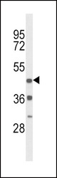
- Experimental details
- Western blot analysis of DRD4 Antibody (Center) (Cat. #AP8760c) in mouse heart tissue lysates (35ug/lane). DRD4 (arrow) was detected using the purified Pab.
- Primary Ab dilution
- 1:1000

Supportive validation
- Submitted by
- Abcepta (provider)
- Main image
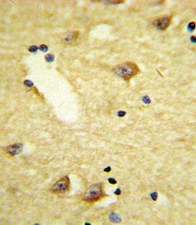
- Experimental details
- "Formalin-fixed and paraffin-embedded human brain tissue reacted with DRD4 Antibody (Center), which was peroxidase-conjugated to the secondary antibody, followed by DAB staining. This data demonstrates the use of this antibody for immunohistochemistry; clinical relevance has not been evaluated."
- Primary Ab dilution
- 1:10~50

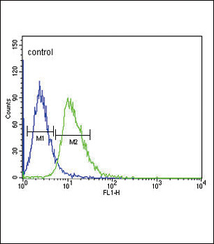
Supportive validation
- Submitted by
- Abcepta (provider)
- Main image
- Experimental details
- DRD4 Antibody (Center) (Cat. #AP8760c) flow cytometric analysis of CEM cells (right histogram) compared to a negative control cell (left histogram).FITC-conjugated goat-anti-rabbit secondary antibodies were used for the analysis.
- Primary Ab dilution
- 1:10~50