Explore
 Validate
 Learn
Learn Western blot
 Immunocytochemistry
Antibody data
- Antibody Data
- Antigen structure
- References [6]
- Comments [0]
- Validations
- Western blot [1]
- Immunoprecipitation [1]
- Immunohistochemistry [1]
Submit
Validation data
Reference
Comment
Report error
- Product number
- NB100-1921 - Provider product page

- Provider
- Novus Biologicals
- Proper citation
- Novus Cat#NB100-1921, RRID:AB_10001061
- Product name
- Rabbit Polyclonal PDI Antibody
- Antibody type
- Polyclonal
- Description
- Unpurified.
- Reactivity
- Human, Mouse, Rat, Bovine, Simian
- Host
- Rabbit
- Isotype
- IgG
- Vial size
- 0.1 ml
- Storage
- Aliquot and store at -20C or -80C. Avoid freeze-thaw cycles.
Submitted references Mitochondrial protein import is regulated by p17/PERMIT to mediate lipid metabolism and cellular stress.
A Split-Luciferase-Based Trimer Formation Assay as a High-throughput Screening Platform for Therapeutics in Alport Syndrome.
GLUT10-Lacking in Arterial Tortuosity Syndrome-Is Localized to the Endoplasmic Reticulum of Human Fibroblasts.
G-CSF improves murine G6PC3-deficient neutrophil function by modulating apoptosis and energy homeostasis.
Mutation in the DC-SIGN cytoplasmic triacidic cluster motif markedly attenuates receptor activity for phagocytosis and endocytosis of mannose-containing ligands by human myeloid cells.
Thyroid hormone-dependent redistribution of the 55-kilodalton monomer of protein disulfide isomerase in cultured glial cells.
Oleinik N, Kim J, Roth BM, Selvam SP, Gooz M, Johnson RH, Lemasters JJ, Ogretmen B
Science advances 2019 Sep;5(9):eaax1978
Science advances 2019 Sep;5(9):eaax1978
A Split-Luciferase-Based Trimer Formation Assay as a High-throughput Screening Platform for Therapeutics in Alport Syndrome.
Omachi K, Kamura M, Teramoto K, Kojima H, Yokota T, Kaseda S, Kuwazuru J, Fukuda R, Koyama K, Matsuyama S, Motomura K, Shuto T, Suico MA, Kai H
Cell chemical biology 2018 May 17;25(5):634-643.e4
Cell chemical biology 2018 May 17;25(5):634-643.e4
GLUT10-Lacking in Arterial Tortuosity Syndrome-Is Localized to the Endoplasmic Reticulum of Human Fibroblasts.
Gamberucci A, Marcolongo P, Németh CE, Zoppi N, Szarka A, Chiarelli N, Hegedűs T, Ritelli M, Carini G, Willaert A, Callewaert BL, Coucke PJ, Benedetti A, Margittai É, Fulceri R, Bánhegyi G, Colombi M
International journal of molecular sciences 2017 Aug 22;18(8)
International journal of molecular sciences 2017 Aug 22;18(8)
G-CSF improves murine G6PC3-deficient neutrophil function by modulating apoptosis and energy homeostasis.
Jun HS, Lee YM, Song KD, Mansfield BC, Chou JY
Blood 2011 Apr 7;117(14):3881-92
Blood 2011 Apr 7;117(14):3881-92
Mutation in the DC-SIGN cytoplasmic triacidic cluster motif markedly attenuates receptor activity for phagocytosis and endocytosis of mannose-containing ligands by human myeloid cells.
Azad AK, Torrelles JB, Schlesinger LS
Journal of leukocyte biology 2008 Dec;84(6):1594-603
Journal of leukocyte biology 2008 Dec;84(6):1594-603
Thyroid hormone-dependent redistribution of the 55-kilodalton monomer of protein disulfide isomerase in cultured glial cells.
Safran M, Farwell AP, Leonard JL
Endocrinology 1992 Nov;131(5):2413-8
Endocrinology 1992 Nov;131(5):2413-8
No comments: Submit comment
Supportive validation
- Submitted by
- Novus Biologicals (provider)
- Main image
- Experimental details
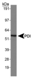
- Western Blot: PDI Antibody [NB100-1921] - Normal human brain.

Supportive validation
- Submitted by
- Novus Biologicals (provider)
- Main image

- Experimental details
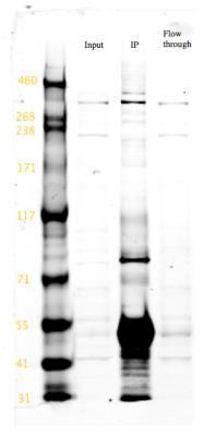
- Immunoprecipitation: PDI Antibody [NB100-1921] - analysis of WDFY3 in E13.5 mouse embryos using anti-WDFY3 antibody. Image from verified customer review.
Supportive validation
- Submitted by
- Novus Biologicals (provider)
- Main image
- Experimental details
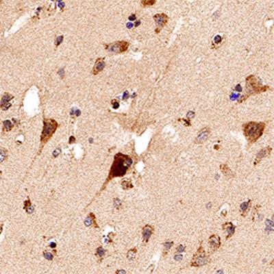
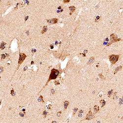
- Immunohistochemistry-Paraffin: PDI Antibody [NB100-1921] - IHC analysis of a formalin fixed paraffin-embedded (FFPE) human brain using 1:250 conc. of PDI antibody on a Bond Rx autostainer (Leica Biosystems). The assay involved 20 minutes of heat induced antigen retrieval (HIER) using 10mM sodium citrate buffer (pH 6.0) and endogenous peroxidase quenching with peroxide block. The sections were incubated with primary antibody for 30 minutes and Bond Polymer Refine Detection (Leica Biosystems) with DAB was used for signal development followed by counterstaining with hematoxylin. Whole slide scanning and capturing of representative images was performed using Aperio AT2 (Leica Biosystems). Cytoplasmic staining of PDI in neurons was observed. Staining was performed by Histowiz.