Explore
 Validate
 Learn
Learn Western blot
 Immunocytochemistry
Antibody data
- Antibody Data
- Antigen structure
- References [1]
- Comments [0]
- Validations
- Immunocytochemistry [1]
Submit
Validation data
Reference
Comment
Report error
- Product number
- MAB2240 - Provider product page

- Provider
- R&D Systems
- Product name
- Human Cadherin-12 Antibody
- Antibody type
- Monoclonal
- Description
- Protein A or G purified from hybridoma culture supernatant. Detects human Cadherin-12 in direct ELISAs and Western blots. In direct ELISAs and Western blots, no cross-reactivity with recombinant human (rh) Cadherin-8, -11, -13, -17, rhE-Cadherin, rhP-Cadherin, rhR-Cadherin, or rhVR-Cadherin is observed.
- Reactivity
- Human
- Host
- Rat
- Conjugate
- Unconjugated
- Antigen sequence
P55289- Isotype
- IgG
- Antibody clone number
- 343621
- Vial size
- 100 ug
- Concentration
- LYOPH
- Storage
- Use a manual defrost freezer and avoid repeated freeze-thaw cycles. 12 months from date of receipt, -20 to -70 °C as supplied. 1 month, 2 to 8 °C under sterile conditions after reconstitution. 6 months, -20 to -70 °C under sterile conditions after reconstitution.
Submitted references N-cadherin is regulated by activin A and associated with tumor aggressiveness in esophageal carcinoma.
Yoshinaga K, Inoue H, Utsunomiya T, Sonoda H, Masuda T, Mimori K, Tanaka Y, Mori M
Clinical cancer research : an official journal of the American Association for Cancer Research 2004 Sep 1;10(17):5702-7
Clinical cancer research : an official journal of the American Association for Cancer Research 2004 Sep 1;10(17):5702-7
No comments: Submit comment
Supportive validation
- Submitted by
- R&D Systems (provider)
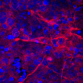
- Main image

- Experimental details
- Cadherin-12 in BG01V Human Embryonic Stem Cells. Cadherin-12 was detected in immersion fixed BG01V human embryonic stem cells differentiated into neurons using Rat Anti-Human Cadherin-12 Monoclonal Antibody (Catalog # MAB2240) at 10 µg/mL for 3 hours at room temperature. Cells were stained using the NorthernLights™ 557-conjugated Anti-Rat IgG Secondary Antibody (red; Catalog # NL013) and counterstained with DAPI (blue). Specific staining was localized to cell membranes. View our protocol for Fluorescent ICC Staining of Stem Cells on Coverslips.