 Explore
 Validate
 Learn
Learn Western blot
 Immunohistochemistry
Antibody data
- Antibody Data
- Antigen structure
- References [1]
- Comments [0]
- Validations
- Immunohistochemistry [1]
- Other assay [1]
Submit
Validation data
Reference
Comment
Report error
- Product number
- PA5-27614 - Provider product page

- Provider
- Invitrogen Antibodies
- Product name
- TALDO1 Polyclonal Antibody
- Antibody type
- Polyclonal
- Antigen
- Recombinant full-length protein
- Description
- Recommended positive controls: A431, mouse brain. Predicted reactivity: Mouse (96%), Rat (96%), Xenopus laevis (80%), Pig (91%), Chicken (82%), Bovine (96%). Store product as a concentrated solution. Centrifuge briefly prior to opening the vial.
- Reactivity
- Human, Mouse, Rat
- Host
- Rabbit
- Isotype
- IgG
- Vial size
- 100 μL
- Concentration
- 6.48 mg/mL
- Storage
- Store at 4°C short term. For long term storage, store at -20°C, avoiding freeze/thaw cycles.
Submitted references The effects of fructose and metabolic inhibition on hepatocellular carcinoma.
Dewdney B, Alanazy M, Gillman R, Walker S, Wankell M, Qiao L, George J, Roberts A, Hebbard L
Scientific reports 2020 Oct 7;10(1):16769
Scientific reports 2020 Oct 7;10(1):16769
No comments: Submit comment
Supportive validation
- Submitted by
- Invitrogen Antibodies (provider)
- Main image
- Experimental details
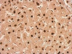
- TALDO1 Polyclonal Antibody detects TALDO1 protein at nucleus on human hepatoma by immunohistochemical analysis. Sample: Paraffin-embedded hepatoma. TALDO1 Polyclonal Antibody (Product # PA5-27614) dilution: 1:500. Antigen Retrieval: EDTA based buffer, pH 8.0, 15 min.
Supportive validation
- Submitted by
- Invitrogen Antibodies (provider)
- Main image
- Experimental details
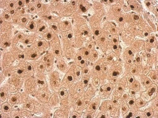
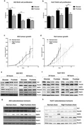
- Figure 1 Evaluating A52 and Huh7 HCC growth and protein expression in glucose and fructose conditions. Cell proliferation via a BrdU assay was determined for ( a ) A52 cells, and ( b ) Huh7 cells grown in media containing 5 mM glucose or 5 mM fructose over 24, 48 and 72 h. Growth of ( c ) A52 and ( d ) Huh7 tumours was evaluated in mice fed a normal or high-fructose chow. Western blots of cell lysates for TKT, TALDO, PHGDH, G6PD, PSAT1 and beta-actin from, ( e ) A52 cells, and ( f ) Huh7 cells growth in glucose or fructose containing media for 24 and 48 h, and tumour lysates from ( g ) A52, and ( h ) Huh7 (H) subcutaneous tumours in mice fed a normal chow or high fructose diet. Original blots and densitometry analysis for the in vitro protein analysis and in vivo protein analysis are shown in Supplementary Figures 1 - 8 (Supplementary File 1 ). Full length western blot images for TKT, TALDO, PHGDH, PSAT1 and beta-actin could not be provided for ( e ) and ( g ). These images were provided from the thesis of M.A., and full-length original images could not be obtained. The original images in this figure and the Supplementary File are as presented in the thesis of M.A. *p < 0.05; **p < 0.01; ***p < 0.001; ****p < 0.0001. G6PD glucose-6-phosphate dehydrogenase, PHGDH phosphoglycerate dehydrogenase, PSAT1 phosphoserine aminotransferase1, TALDO transaldolase, TKT transketolase.
