Explore
 Validate
 Learn
LearnALX-804-837-C100
antibody from Enzo Life Sciences
Targeting: PIDD1
DKFZp434D229, LRDD, MGC16925, PIDD
 Western blot
 Immunocytochemistry
Antibody data
- Antibody Data
- Antigen structure
- References [3]
- Comments [0]
- Validations
- Immunocytochemistry [2]
Submit
Validation data
Reference
Comment
Report error
- Product number
- ALX-804-837-C100 - Provider product page

- Provider
- Enzo Life Sciences
- Proper citation
- Enzo Life Sciences Cat#ALX-804-837-C100, RRID:AB_2052254
- Product name
- PIDD monoclonal antibody (Anto-1)
- Antibody type
- Monoclonal
- Antigen
- Recombinant protein fragment
- Reactivity
- Human, Mouse
- Host
- Mouse
- Isotype
- IgG
- Antibody clone number
- Anto-1
- Vial size
- 100 μg
- Storage
- -20°C
- Handling
- Avoid freeze/thaw cycles. After opening, prepare aliquots and store at -20¡C.
Submitted references Caspase-2 is involved in cell death induction by taxanes in breast cancer cells.
Intrinsic caspase-8 activation mediates sensitization of erlotinib-resistant tumor cells to erlotinib/cell-cycle inhibitors combination treatment.
Autoproteolysis of PIDD marks the bifurcation between pro-death caspase-2 and pro-survival NF-kappaB pathway.
Jelínek M, Balušíková K, Kopperová D, Nĕmcová-Fürstová V, Šrámek J, Fidlerová J, Zanardi I, Ojima I, Kovář J
Cancer cell international 2013 May 15;13(1):42
Cancer cell international 2013 May 15;13(1):42
Intrinsic caspase-8 activation mediates sensitization of erlotinib-resistant tumor cells to erlotinib/cell-cycle inhibitors combination treatment.
Orzáez M, Guevara T, Sancho M, Pérez-Payá E
Cell death & disease 2012 Oct 25;3:e415
Cell death & disease 2012 Oct 25;3:e415
Autoproteolysis of PIDD marks the bifurcation between pro-death caspase-2 and pro-survival NF-kappaB pathway.
Tinel A, Janssens S, Lippens S, Cuenin S, Logette E, Jaccard B, Quadroni M, Tschopp J
The EMBO journal 2007 Jan 10;26(1):197-208
The EMBO journal 2007 Jan 10;26(1):197-208
No comments: Submit comment
Supportive validation
- Submitted by
- Enzo Life Sciences (provider)
- Main image

- Experimental details
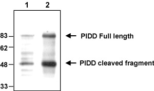
- Western blot analysis of endogenous and overexpressed human PIDD usingÊMAb to PIDD (Anto-1) (Prod. No. ALX-804-837). Method: Cell extracts (1x10E5) from HEK 293T wild type (lane 1) or transfected with a plasmid coding for human PIDD (lane 2) were resolved by SDS-PAGE under reducing conditions, transferred to nitrocellulose and incubated with the MAb to PIDDÊ(Anto-1) at a 1:500 dilution for 2 hours. Proteins were visualized using a peroxidase-conjugated polyclonal antibody to mouse IgG and a chemiluminescence detection system. Note that two major forms of PIDD are detected, representing full length and cleaved proteins, migrating at approximatively 90kDa and 50kDa, respectively.
- Submitted by
- Enzo Life Sciences (provider)
- Main image

- Experimental details
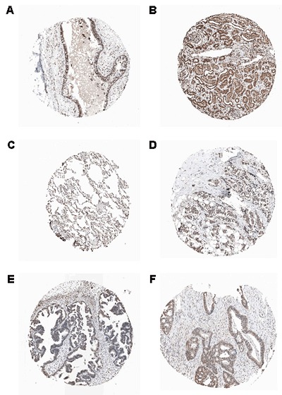
- Immunohistochemical staining of endogenous human PIDD in different human tissues (paraffin sections) using MAb to PIDD (Anto-1) (Prod. No. ALX-804-837). Method: Different human normal (A: Bronchus; B: Kidney; C: Lung) or cancer (D: Breast; E: Ovarian; F: Colo-rectal) tissues were stained with MAb to PIDD (Anto-1) by standard immunohistochemistry. Pictures courtesy of Human Protein Atlas (www.proteinatlas.org).