 Explore
 Validate
 Learn
Learn Flow cytometry
Antibody data
- Antibody Data
- Antigen structure
- References [1]
- Comments [0]
- Validations
- Flow cytometry [2]
Submit
Validation data
Reference
Comment
Report error
- Product number
- FAB23193N - Provider product page

- Provider
- Novus Biologicals
- Product name
- Mouse Monoclonal LAG-3 Antibody
- Antibody type
- Monoclonal
- Description
- Protein A or G purified from hybridoma culture supernatant. Detects human LAG-3 in direct ELISAs.
- Reactivity
- Human
- Host
- Mouse
- Conjugate
- Near infrared dye
- Isotype
- IgG
- Vial size
- 100 Tests
- Storage
- Protect from light. Do not freeze. 12 months from date of receipt, 2 to 8 degreesC as supplied.
Submitted references PD-1(hi)TIM-3(+) T cells associate with and predict leukemia relapse in AML patients post allogeneic stem cell transplantation.
Kong Y, Zhang J, Claxton DF, Ehmann WC, Rybka WB, Zhu L, Zeng H, Schell TD, Zheng H
Blood cancer journal 2015 Jul 31;5:e330
Blood cancer journal 2015 Jul 31;5:e330
No comments: Submit comment
Supportive validation
- Submitted by
- Novus Biologicals (provider)
- Main image
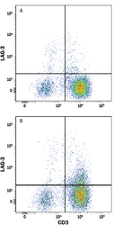
- Experimental details
- Detection of LAG-3 in Human PBMCs by Flow Cytometry. Human peripheral blood mononuclear cells (PBMCs) either (A) untreated or (B) treated with 1 μg/mL PHA for 5 days were stained with Mouse Anti-Human LAG-3 Alexa Fluor® 700-conjugated Monoclonal Antibody (Catalog # FAB23193N) and Mouse Anti-Human CD3 epsilon PE-conjugated Monoclonal Antibody (Catalog # FAB100P). Quadrant markers were set based on control antibody staining (Catalog # IC002N). View our protocol for Staining Membrane-associated Proteins.

- Submitted by
- Novus Biologicals (provider)
- Main image

- Experimental details
- Detection of LAG-3 in HEK293 Human Cell Line Transfected with Human LAG-3 and eGFP by Flow Cytometry. HEK293 human embryonic kidney cell line transfected with either (A) human LAG-3 or (B) irrelelvant transfectants and eGFP was stained with Mouse Anti-Human LAG-3 Alexa Fluor® 700-conjugated Monoclonal Antibody (Catalog # FAB23193N). Quadrant markers were set based on control antibody staining (Catalog # IC002N, data not shown). View our protocol for Staining Membrane-associated Proteins.
