 Explore
 Validate
 Learn
Learn Western blot
Antibody data
- Antibody Data
- Antigen structure
- References [1]
- Comments [0]
- Validations
- Western blot [1]
Submit
Validation data
Reference
Comment
Report error
- Product number
- AF3328 - Provider product page

- Provider
- R&D Systems
- Product name
- Mouse LAG-3 Antibody
- Antibody type
- Polyclonal
- Description
- Antigen Affinity-purified. Detects mouse LAG-3 in direct ELISAs and Western blots. In direct ELISAs and Western blots, less than 1% cross-reactivity with recombinant human LAG-3 is observed.
- Reactivity
- Mouse
- Host
- Goat
- Conjugate
- Unconjugated
- Antigen sequence
Q61790- Isotype
- IgG
- Vial size
- 100 ug
- Concentration
- LYOPH
- Storage
- Use a manual defrost freezer and avoid repeated freeze-thaw cycles. 12 months from date of receipt, -20 to -70 °C as supplied. 1 month, 2 to 8 °C under sterile conditions after reconstitution. 6 months, -20 to -70 °C under sterile conditions after reconstitution.
Submitted references Modulation of redox balance leaves murine diabetogenic TH1 T cells "LAG-3-ing" behind.
Delmastro MM, Styche AJ, Trucco MM, Workman CJ, Vignali DA, Piganelli JD
Diabetes 2012 Jul;61(7):1760-8
Diabetes 2012 Jul;61(7):1760-8
No comments: Submit comment
Supportive validation
- Submitted by
- R&D Systems (provider)
- Main image
- Experimental details
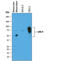
- Detection of Mouse LAG-3 by Western Blot. Western blot shows lysates of mouse liver tissue, mouse heart tissue, EL4.IL-2 mouse lymphoblast cell line, and CTLL-2 mouse cytotoxic T cell line. PVDF membrane was probed with 0.25 µg/mL of Goat Anti-Mouse LAG-3 Antigen Affinity-purified Polyclonal Antibody (Catalog # AF3328) followed by HRP-conjugated Anti-Goat IgG Secondary Antibody (Catalog # HAF019). Specific bands were detected for LAG-3 at approximately 54 kDa and 75 kDa in mouse liver and mouse heart tissue and 70-80 kDa in EL4.IL-2 and CTLL-2 cell lines (as indicated). This experiment was conducted under reducing conditions and using Immunoblot Buffer Group 1.
