Explore
 Validate
 Learn
Learn Flow cytometry
Antibody data
- Antibody Data
- Antigen structure
- References [3]
- Comments [0]
- Validations
- Flow cytometry [2]
- Other assay [4]
Submit
Validation data
Reference
Comment
Report error
- Product number
- 62-2239-41 - Provider product page

- Provider
- Invitrogen Antibodies
- Product name
- CD223 (LAG-3) Monoclonal Antibody (3DS223H), Super Bright™ 436, eBioscience™
- Antibody type
- Monoclonal
- Antigen
- Other
- Description
- Description: This 3DS223H monoclonal antibody recognizes human CD223 also known as Lymphocyte Activation Gene 3 (LAG-3). LAG-3 is a 70-kDa surface glycoprotein belonging to the Ig superfamily with homology to CD4. LAG-3 binds to MHC class II with higher affinity than CD4 and is thought to be involved in the negative regulation of T cell activation and homeostatic proliferation. Surface expression of LAG-3 has been reported on activated T cells (including regulatory T cells) and NK cells. CD8+ T cells usually express LAG-3 at significantly higher levels than CD4+ T cells. Coexpression of LAG-3 and CD49b has been proposed to identify human and mouse Type 1 regulatory T cells (Tr1 cells). This 3DS223H antibody will recognize a formaldehyde-fixed epitope. Applications Reported: This 3DS223H antibody has been reported for use in flow cytometric analysis. Applications Tested: This 3DS223H antibody has been pre-titrated and tested by flow cytometric analysis of stimulated normal human peripheral blood cells. This can be used at 5 µL (0.125 µg) per test. A test is defined as the amount (µg) of antibody that will stain a cell sample in a final volume of 100 µL. Cell number should be determined empirically but can range from 10^5 to 10^8 cells/test. Super Bright 436 can be excited with the violet laser line (405 nm) and emits at 436 nm. We recommend using a 450/50 bandpass filter, or equivalent. Please make sure that your instrument is capable of detecting this fluorochrome. When using two or more Super Bright dye-conjugated antibodies in a staining panel, it is recommended to use Super Bright Complete Staining Buffer (Product # SB-4401) to minimize any non-specific polymer interactions. Please refer to the datasheet for Super Bright Staining Buffer for more information. Excitation: 405 nm; Emission: 436 nm; Laser: Violet Laser Super Bright Polymer Dyes are sold under license from Becton, Dickinson and Company.
- Reactivity
- Human
- Host
- Mouse
- Isotype
- IgG
- Antibody clone number
- 3DS223H
- Vial size
- 25 Tests
- Concentration
- 5 μL/Test
- Storage
- 4°C, store in dark, DO NOT FREEZE!
Submitted references MAVS Genetic Variation Is Associated with Decreased HIV-1 Replication In Vitro and Reduced CD4(+) T Cell Infection in HIV-1-Infected Individuals.
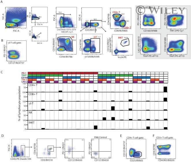
OMIP-037: 16-color panel to measure inhibitory receptor signatures from multiple human immune cell subsets.
Follicular regulatory T cells impair follicular T helper cells in HIV and SIV infection.
Stunnenberg M, van Pul L, Sprokholt JK, van Dort KA, Gringhuis SI, Geijtenbeek TBH, Kootstra NA
Viruses 2020 Jul 16;12(7)
Viruses 2020 Jul 16;12(7)
OMIP-037: 16-color panel to measure inhibitory receptor signatures from multiple human immune cell subsets.
Belkina AC, Snyder-Cappione JE
Cytometry. Part A : the journal of the International Society for Analytical Cytology 2017 Feb;91(2):175-179
Cytometry. Part A : the journal of the International Society for Analytical Cytology 2017 Feb;91(2):175-179
Follicular regulatory T cells impair follicular T helper cells in HIV and SIV infection.
Miles B, Miller SM, Folkvord JM, Kimball A, Chamanian M, Meditz AL, Arends T, McCarter MD, Levy DN, Rakasz EG, Skinner PJ, Connick E
Nature communications 2015 Oct 20;6:8608
Nature communications 2015 Oct 20;6:8608
No comments: Submit comment
Supportive validation
- Submitted by
- Invitrogen Antibodies (provider)
- Main image
- Experimental details
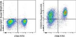
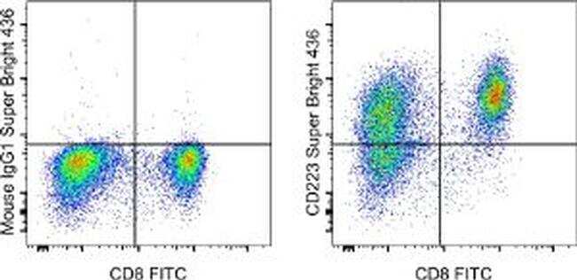
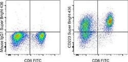
- Staining of 3-day Anti-Human CD3 and CD28 Functional Grade Purified (Product # 16-0037-81 and Product # 16-0289-81)-stimulated normal human peripheral blood cells with Anti-Human CD8a FITC (Product # 11-0087-42) and Mouse IgG1 K Isotype Control Super Bright 436 (Product # 62-4714-82) (left) or Anti-Human CD223 (LAG-3) Super Bright 436 (right). Viable cells in the lymphocyte gate, as determined by 7-AAD Viability Staining Solution (Product # 00-6993-50) were used for analysis.
- Submitted by
- Invitrogen Antibodies (provider)
- Main image
- Experimental details
- Staining of 3-day Anti-Human CD3 and CD28 Functional Grade Purified (Product # 16-0037-81 and Product # 16-0289-81)-stimulated normal human peripheral blood cells with Anti-Human CD8a FITC (Product # 11-0087-42) and Mouse IgG1 K Isotype Control Super Bright 436 (Product # 62-4714-82) (left) or Anti-Human CD223 (LAG-3) Super Bright 436 (right). Viable cells in the lymphocyte gate, as determined by 7-AAD Viability Staining Solution (Product # 00-6993-50) were used for analysis.

Supportive validation
- Submitted by
- Invitrogen Antibodies (provider)
- Main image

- Experimental details
- NULL
- Submitted by
- Invitrogen Antibodies (provider)
- Main image

- Experimental details
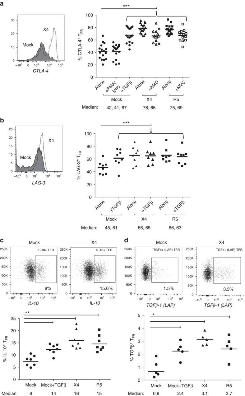
- Figure 6 T FR exhibit an enhanced regulatory phenotype in ex vivo HIV infection. Tonsil cells were mock-, X4-, or R5-spinoculated and cultured under experimental conditions as indicated. T FR were then analysed for expression of regulatory receptors and cytokine production by intracellular cytokine staining. ( a ) Percentage of total (surface and intracellular) T FR CTLA-4 expression ( n =15). ( b ) Percentage of surface T FR LAG-3 expression ( n =8). ( c ) Production of IL-10 by T FR ( n =7). ( d ) Production of TGF-beta-1 (measured as LAP) by T FR ( n =5). The horizontal bars of each graph indicate the median value and are listed where appropriate for clarity. Statistical analyses were performed by Friedman ( a , b ) or Mann-Whitney tests ( c , d ) and significance is denoted by asterisks where * P
- Submitted by
- Invitrogen Antibodies (provider)
- Main image
- Experimental details
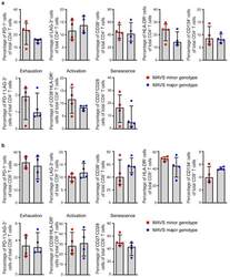
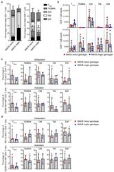
- Figure 3 MAVS genetic variation does not affect T cell exhaustion, activation, and senescence. ( a ) Percentages of PD-1 + , LAG-3 + , CD38 + , HLA-DR + , CD134 + , exhausted (PD-1 + LAG-3 + ), activated (CD38 + HLA-DR + ), and senescent (CD27 - CD28 - ) cells within CD4 + and ( b ) CD8 + T cells of untreated HIV-1-infected individuals with a MAVS minor or MAVS major genotype 2.5-3.5 years p.SC were analyzed using flow cytometry. Each square or dot represents a different study participant (median +- IQR). No significant differences between HIV-1-infected individuals with a MAVS minor or MAVS major genotype were observed.
- Submitted by
- Invitrogen Antibodies (provider)
- Main image
- Experimental details
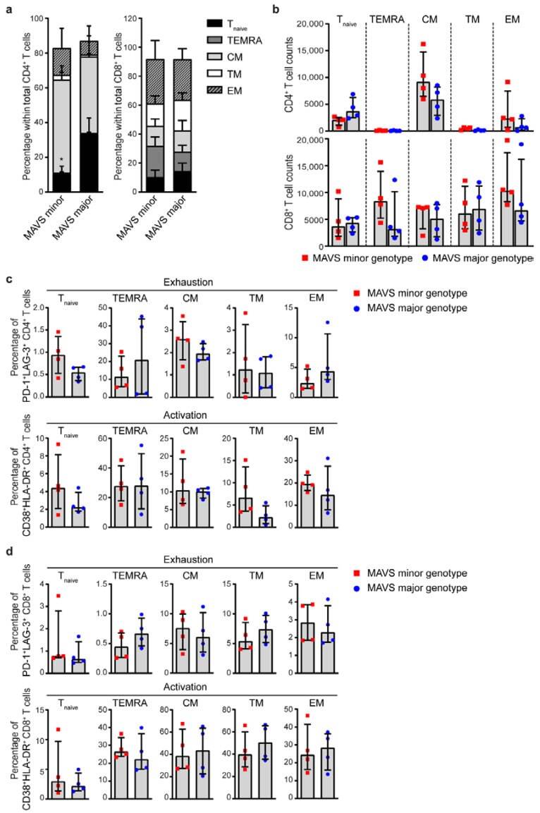
- Figure 4 MAVS minor genotype is associated with a decreased percentage of naive CD4 + T cells. ( a ) Percentages and ( b ) cell counts of naive (T naive ; CD45RA + CD27 + CCR7 + ), terminally differentiated effector memory (TEMRA; CD45RA + CCR7 - CD27 - ), central memory (CM; CD45RA - CCR7 + CD27 + ), transitional memory (TM; CD45RA - CCR7 - CD27 + ), and effector memory (EM; CD45RA - CCR7 - CD27 - ) cells within CD4 + and CD8 + T cells were analyzed using flow cytometry. ( c ) Percentages of exhausted (PD-1 + LAG-3 + ) and activated (CD38 + HLA-DR + ) CD4 + T cells and ( d ) CD8 + T cells within T naive , TEMRA, CM, TM, and EM populations were analyzed using flow cytometry. Each square or dot represents a different study participant (median +- IQR). All significant differences are indicated: * p < 0.05, unpaired Mann-Whitney test.