Explore
 Validate
 Learn
Learn Immunohistochemistry
 Flow cytometry
Antibody data
- Antibody Data
- Antigen structure
- References [16]
- Comments [0]
- Validations
- Flow cytometry [5]
Submit
Validation data
Reference
Comment
Report error
- Product number
- NBP1-97665 - Provider product page

- Provider
- Novus Biologicals
- Proper citation
- Novus Cat#NBP1-97665, RRID:AB_11190252
- Product name
- Mouse Monoclonal LAG-3 Antibody
- Antibody type
- Monoclonal
- Description
- Protein A or G purified. Recognizes the 30 aa extra-loop of the first N-terminal D1 domain of human LAG-3.
- Reactivity
- Human, Simian
- Host
- Mouse
- Conjugate
- Green dye
- Isotype
- IgG
- Vial size
- 0.05 mg
- Storage
- Store at 4C in the dark.
Submitted references CTLA-4 and PD-1 dual blockade induces SIV reactivation without control of rebound after antiretroviral therapy interruption.
Profiling Immune Escape in Hodgkin's and Diffuse large B-Cell Lymphomas Using the Transcriptome and Immunostaining.
Human lymphocyte activation gene-3 molecules expressed by activated T cells deliver costimulation signal for dendritic cell activation.
Human lymphocyte activation gene-3 molecules expressed by activated T cells deliver costimulation signal for dendritic cell activation.
A soluble lymphocyte activation gene-3 (sLAG-3) protein as a prognostic factor in human breast cancer expressing estrogen or progesterone receptors.
The negative regulatory function of the lymphocyte-activation gene-3 co-receptor (CD223) on human T cells.
Immunological mechanisms elicited at the tumour site by lymphocyte activation gene-3 (LAG-3) versus IL-12: sharing a common Th1 anti-tumour immune pathway.
T Lymphocytes infiltrating various tumour types express the MHC class II ligand lymphocyte activation gene-3 (LAG-3): role of LAG-3/MHC class II interactions in cell-cell contacts.
T Lymphocytes infiltrating various tumour types express the MHC class II ligand lymphocyte activation gene-3 (LAG-3): role of LAG-3/MHC class II interactions in cell-cell contacts.
CD3/TCR complex-associated lymphocyte activation gene-3 molecules inhibit CD3/TCR signaling.
Characterization of the major histocompatibility complex class II binding site on LAG-3 protein.
Characterization of the major histocompatibility complex class II binding site on LAG-3 protein.
Cellular expression and tissue distribution of the human LAG-3-encoded protein, an MHC class II ligand.
Cellular expression and tissue distribution of the human LAG-3-encoded protein, an MHC class II ligand.
Characterization of the lymphocyte activation gene 3-encoded protein. A new ligand for human leukocyte antigen class II antigens.
Characterization of the lymphocyte activation gene 3-encoded protein. A new ligand for human leukocyte antigen class II antigens.
Harper J, Gordon S, Chan CN, Wang H, Lindemuth E, Galardi C, Falcinelli SD, Raines SLM, Read JL, Nguyen K, McGary CS, Nekorchuk M, Busman-Sahay K, Schawalder J, King C, Pino M, Micci L, Cervasi B, Jean S, Sanderson A, Johns B, Koblansky AA, Amrine-Madsen H, Lifson J, Margolis DM, Silvestri G, Bar KJ, Favre D, Estes JD, Paiardini M
Nature medicine 2020 Apr;26(4):519-528
Nature medicine 2020 Apr;26(4):519-528
Profiling Immune Escape in Hodgkin's and Diffuse large B-Cell Lymphomas Using the Transcriptome and Immunostaining.
Péricart S, Tosolini M, Gravelle P, Rossi C, Traverse-Glehen A, Amara N, Franchet C, Martin E, Bezombes C, Laurent G, Brousset P, Fournié JJ, Laurent C
Cancers 2018 Oct 31;10(11)
Cancers 2018 Oct 31;10(11)
Human lymphocyte activation gene-3 molecules expressed by activated T cells deliver costimulation signal for dendritic cell activation.
Casati C, Camisaschi C, Novellino L, Mazzocchi A, Triebel F, Rivoltini L, Parmiani G, Castelli C
Journal of immunology (Baltimore, Md. : 1950) 2008 Mar 15;180(6):3782-8
Journal of immunology (Baltimore, Md. : 1950) 2008 Mar 15;180(6):3782-8
A soluble lymphocyte activation gene-3 (sLAG-3) protein as a prognostic factor in human breast cancer expressing estrogen or progesterone receptors.
Triebel F, Hacene K, Pichon MF
Cancer letters 2006 Apr 8;235(1):147-53
Cancer letters 2006 Apr 8;235(1):147-53
The negative regulatory function of the lymphocyte-activation gene-3 co-receptor (CD223) on human T cells.
Maçon-Lemaître L, Triebel F
Immunology 2005 Jun;115(2):170-8
Immunology 2005 Jun;115(2):170-8
Immunological mechanisms elicited at the tumour site by lymphocyte activation gene-3 (LAG-3) versus IL-12: sharing a common Th1 anti-tumour immune pathway.
Di Carlo E, Cappello P, Sorrentino C, D'Antuono T, Pellicciotta A, Giovarelli M, Forni G, Musiani P, Triebel F
The Journal of pathology 2005 Jan;205(1):82-91
The Journal of pathology 2005 Jan;205(1):82-91
T Lymphocytes infiltrating various tumour types express the MHC class II ligand lymphocyte activation gene-3 (LAG-3): role of LAG-3/MHC class II interactions in cell-cell contacts.
Demeure CE, Wolfers J, Martin-Garcia N, Gaulard P, Triebel F
European journal of cancer (Oxford, England : 1990) 2001 Sep;37(13):1709-18
European journal of cancer (Oxford, England : 1990) 2001 Sep;37(13):1709-18
CD3/TCR complex-associated lymphocyte activation gene-3 molecules inhibit CD3/TCR signaling.
Hannier S, Tournier M, Bismuth G, Triebel F
Journal of immunology (Baltimore, Md. : 1950) 1998 Oct 15;161(8):4058-65
Journal of immunology (Baltimore, Md. : 1950) 1998 Oct 15;161(8):4058-65
Characterization of the major histocompatibility complex class II binding site on LAG-3 protein.
Huard B, Mastrangeli R, Prigent P, Bruniquel D, Donini S, El-Tayar N, Maigret B, Dréano M, Triebel F
Proceedings of the National Academy of Sciences of the United States of America 1997 May 27;94(11):5744-9
Proceedings of the National Academy of Sciences of the United States of America 1997 May 27;94(11):5744-9
Cellular expression and tissue distribution of the human LAG-3-encoded protein, an MHC class II ligand.
Huard B, Gaulard P, Faure F, Hercend T, Triebel F
Immunogenetics 1994;39(3):213-7
Immunogenetics 1994;39(3):213-7
Cellular expression and tissue distribution of the human LAG-3-encoded protein, an MHC class II ligand.
Huard B, Gaulard P, Faure F, Hercend T, Triebel F
Immunogenetics 1994;39(3):213-7
Immunogenetics 1994;39(3):213-7
Characterization of the lymphocyte activation gene 3-encoded protein. A new ligand for human leukocyte antigen class II antigens.
Baixeras E, Huard B, Miossec C, Jitsukawa S, Martin M, Hercend T, Auffray C, Triebel F, Piatier-Tonneau D
The Journal of experimental medicine 1992 Aug 1;176(2):327-37
The Journal of experimental medicine 1992 Aug 1;176(2):327-37
No comments: Submit comment
Supportive validation
- Submitted by
- Novus Biologicals (provider)
- Main image

- Experimental details
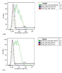
- Flow Cytometry: LAG-3 Antibody (17B4) [FITC] [NBP1-97665] - LAG-3 expression on activated human peripheral blood mononuclear cells (PBMC) detected with LAG-3 (human), mAb (17B4) (FITC).Method: T lymphocytes from human PBMC are stimulated with 1ug/ml of PHA for three days. Then, after seven days of culture, 3x10^6 three-days PHA-activated human PBMC are treated with LAG-3 (human), mAb (17B4) (FITC) or FITC coupled isotype-matched (IgG1) control MAb (used at a saturating dilution of 1:800 and 1:150 respectively) for 30 min. at 4C in RPMI 1640 and washed twice with 1x PBS. Stained cells are then analysed by FC [4].
- Submitted by
- Novus Biologicals (provider)
- Main image

- Experimental details
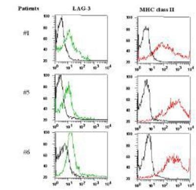
- Flow Cytometry: LAG-3 Antibody (17B4) [FITC] [NBP1-97665] - Tumor infiltrating lymphocytes (TILs) express LAG-3 (detected using LAG-3 (human), mAb (17B4). Method: Freshly dissociated single cell suspensions of renal cell carcinoma TILs are incubated with LAG-3 (human), mAb (17B4) (FITC) (5ug/ml) and anti-MHC Class II molecules (PE) for 30 min. and washed twice in saline buffer. Additional staining with anti-CD3 allowed a gate analysis of total T cells. The LAG-3 and MHC II profiles of CD3+-gated cells for 3 patients are shown [5].
- Submitted by
- Novus Biologicals (provider)
- Main image
- Experimental details
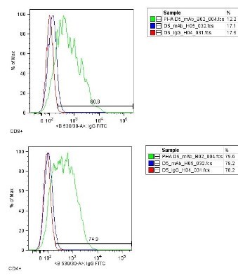
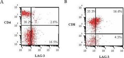
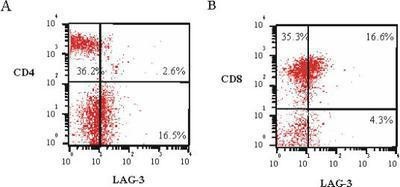
- Flow Cytometry: LAG-3 Antibody (17B4) [FITC] [NBP1-97665] - Expression of LAG-3 on CD4+ and CD8+ subpopulations of tumour infiltrating lymphocytes (TILs) detected with LAG-3 (human), mAb (17B4) (FITC). Method: TILs from a dissociated renal cell carcinoma sample, stained with 5ug/ml LAG-3 (human), mAb (17B4) (FITC) and FITC-coupled anti-CD4 or -CD8, are analyzed by a two-colour FACS analysis. Additional staining with anti-CD3 allowed a gate analysis of total T cells. Values indicate percentages in each quadrant [5].
- Submitted by
- Novus Biologicals (provider)
- Main image
- Experimental details
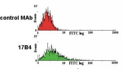
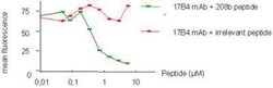
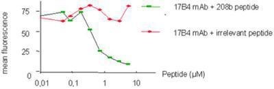
- Flow Cytometry: LAG-3 Antibody (17B4) [FITC] [NBP1-97665] - Specific inhibition of 17B4 staining.Method: LAG-3 (human), mAb (17B4) (FITC) (10ug/ml) (is preincubated with a specific peptide epitope (208b) or a control tetanus toxoid (TT) peptide at different molarities prior to staining of TILs. Stained cells are then analyzed by FC.
- Submitted by
- Novus Biologicals (provider)
- Main image
- Experimental details
- Flow Cytometry: LAG-3 Antibody (17B4) [FITC] [NBP1-97665] - LAG-3 staining in resting and PHA activated lymphocytes. Image from verified customer review.