 Explore
 Validate
 Learn
Learn Immunocytochemistry
 Flow cytometry
Antibody data
- Antibody Data
- Antigen structure
- References [17]
- Comments [0]
- Validations
- Immunocytochemistry [4]
Submit
Validation data
Reference
Comment
Report error
- Product number
- ALX-804-806F-C100 - Provider product page

- Provider
- Enzo Life Sciences
- Product name
- LAG-3 (human) monoclonal antibody (17B4) (FITC conjugate)
- Antibody type
- Monoclonal
- Antigen
- Synthetic peptide
- Description
- Purified from concentrated hybridoma tissue culture supernatant by Protein A Sepharoseª CL-4B Flow Fast Column.
- Reactivity
- Human
- Host
- Mouse
- Conjugate
- Green dye
- Isotype
- IgG
- Antibody clone number
- 17B4
- Vial size
- 100 μg
- Storage
- +4°C
- Handling
- Do not freeze. Protect from light.
Submitted references Elevated expression of LAG-3, but not PD-1, is associated with impaired iNKT cytokine production during chronic HIV-1 infection and treatment.
CD26-mediated induction of EGR2 and IL-10 as potential regulatory mechanism for CD26 costimulatory pathway.
Fusion of ubiquitin to HIV gag impairs human monocyte-derived dendritic cell maturation and reduces ability to induce gag T cell responses.
PD-1 identifies the patient-specific CD8⁺ tumor-reactive repertoire infiltrating human tumors.
Enrichment of LAG-3, but not PD-1, on double negative T cells at the female genital tract.
Selection of CD8+PD-1+ lymphocytes in fresh human melanomas enriches for tumor-reactive T cells.
Human lymphocyte activation gene-3 molecules expressed by activated T cells deliver costimulation signal for dendritic cell activation.
Human dendritic cells acquire a semimature phenotype and lymph node homing potential through interaction with CD4+CD25+ regulatory T cells.
A soluble lymphocyte activation gene-3 (sLAG-3) protein as a prognostic factor in human breast cancer expressing estrogen or progesterone receptors.
Responses to human CD40 ligand/human interleukin-2 autologous cell vaccine in patients with B-cell chronic lymphocytic leukemia.
The negative regulatory function of the lymphocyte-activation gene-3 co-receptor (CD223) on human T cells.
Immunological mechanisms elicited at the tumour site by lymphocyte activation gene-3 (LAG-3) versus IL-12: sharing a common Th1 anti-tumour immune pathway.
T Lymphocytes infiltrating various tumour types express the MHC class II ligand lymphocyte activation gene-3 (LAG-3): role of LAG-3/MHC class II interactions in cell-cell contacts.
CD3/TCR complex-associated lymphocyte activation gene-3 molecules inhibit CD3/TCR signaling.
Characterization of the major histocompatibility complex class II binding site on LAG-3 protein.
Cellular expression and tissue distribution of the human LAG-3-encoded protein, an MHC class II ligand.
Characterization of the lymphocyte activation gene 3-encoded protein. A new ligand for human leukocyte antigen class II antigens.
Juno JA, Stalker AT, Waruk JL, Oyugi J, Kimani M, Plummer FA, Kimani J, Fowke KR
Retrovirology 2015 Feb 13;12:17
Retrovirology 2015 Feb 13;12:17
CD26-mediated induction of EGR2 and IL-10 as potential regulatory mechanism for CD26 costimulatory pathway.
Hatano R, Ohnuma K, Otsuka H, Komiya E, Taki I, Iwata S, Dang NH, Okumura K, Morimoto C
Journal of immunology (Baltimore, Md. : 1950) 2015 Feb 1;194(3):960-72
Journal of immunology (Baltimore, Md. : 1950) 2015 Feb 1;194(3):960-72
Fusion of ubiquitin to HIV gag impairs human monocyte-derived dendritic cell maturation and reduces ability to induce gag T cell responses.
Herath S, Benlahrech A, Papagatsias T, Athanasopoulos T, Bouzeboudjen Z, Hervouet C, Klavinskis L, Meiser A, Kelleher P, Dickson G, Patterson S
PloS one 2014;9(2):e88327
PloS one 2014;9(2):e88327
PD-1 identifies the patient-specific CD8⁺ tumor-reactive repertoire infiltrating human tumors.
Gros A, Robbins PF, Yao X, Li YF, Turcotte S, Tran E, Wunderlich JR, Mixon A, Farid S, Dudley ME, Hanada K, Almeida JR, Darko S, Douek DC, Yang JC, Rosenberg SA
The Journal of clinical investigation 2014 May;124(5):2246-59
The Journal of clinical investigation 2014 May;124(5):2246-59
Enrichment of LAG-3, but not PD-1, on double negative T cells at the female genital tract.
Juno JA, Lajoie J, Stalker AT, Oyugi J, Kimani M, Kimani J, Plummer FA, Fowke KR
American journal of reproductive immunology (New York, N.Y. : 1989) 2014 Dec;72(6):534-40
American journal of reproductive immunology (New York, N.Y. : 1989) 2014 Dec;72(6):534-40
Selection of CD8+PD-1+ lymphocytes in fresh human melanomas enriches for tumor-reactive T cells.
Inozume T, Hanada K, Wang QJ, Ahmadzadeh M, Wunderlich JR, Rosenberg SA, Yang JC
Journal of immunotherapy (Hagerstown, Md. : 1997) 2010 Nov-Dec;33(9):956-64
Journal of immunotherapy (Hagerstown, Md. : 1997) 2010 Nov-Dec;33(9):956-64
Human lymphocyte activation gene-3 molecules expressed by activated T cells deliver costimulation signal for dendritic cell activation.
Casati C, Camisaschi C, Novellino L, Mazzocchi A, Triebel F, Rivoltini L, Parmiani G, Castelli C
Journal of immunology (Baltimore, Md. : 1950) 2008 Mar 15;180(6):3782-8
Journal of immunology (Baltimore, Md. : 1950) 2008 Mar 15;180(6):3782-8
Human dendritic cells acquire a semimature phenotype and lymph node homing potential through interaction with CD4+CD25+ regulatory T cells.
Bayry J, Triebel F, Kaveri SV, Tough DF
Journal of immunology (Baltimore, Md. : 1950) 2007 Apr 1;178(7):4184-93
Journal of immunology (Baltimore, Md. : 1950) 2007 Apr 1;178(7):4184-93
A soluble lymphocyte activation gene-3 (sLAG-3) protein as a prognostic factor in human breast cancer expressing estrogen or progesterone receptors.
Triebel F, Hacene K, Pichon MF
Cancer letters 2006 Apr 8;235(1):147-53
Cancer letters 2006 Apr 8;235(1):147-53
Responses to human CD40 ligand/human interleukin-2 autologous cell vaccine in patients with B-cell chronic lymphocytic leukemia.
Biagi E, Rousseau R, Yvon E, Schwartz M, Dotti G, Foster A, Havlik-Cooper D, Grilley B, Gee A, Baker K, Carrum G, Rice L, Andreeff M, Popat U, Brenner M
Clinical cancer research : an official journal of the American Association for Cancer Research 2005 Oct 1;11(19 Pt 1):6916-23
Clinical cancer research : an official journal of the American Association for Cancer Research 2005 Oct 1;11(19 Pt 1):6916-23
The negative regulatory function of the lymphocyte-activation gene-3 co-receptor (CD223) on human T cells.
Maçon-Lemaître L, Triebel F
Immunology 2005 Jun;115(2):170-8
Immunology 2005 Jun;115(2):170-8
Immunological mechanisms elicited at the tumour site by lymphocyte activation gene-3 (LAG-3) versus IL-12: sharing a common Th1 anti-tumour immune pathway.
Di Carlo E, Cappello P, Sorrentino C, D'Antuono T, Pellicciotta A, Giovarelli M, Forni G, Musiani P, Triebel F
The Journal of pathology 2005 Jan;205(1):82-91
The Journal of pathology 2005 Jan;205(1):82-91
T Lymphocytes infiltrating various tumour types express the MHC class II ligand lymphocyte activation gene-3 (LAG-3): role of LAG-3/MHC class II interactions in cell-cell contacts.
Demeure CE, Wolfers J, Martin-Garcia N, Gaulard P, Triebel F
European journal of cancer (Oxford, England : 1990) 2001 Sep;37(13):1709-18
European journal of cancer (Oxford, England : 1990) 2001 Sep;37(13):1709-18
CD3/TCR complex-associated lymphocyte activation gene-3 molecules inhibit CD3/TCR signaling.
Hannier S, Tournier M, Bismuth G, Triebel F
Journal of immunology (Baltimore, Md. : 1950) 1998 Oct 15;161(8):4058-65
Journal of immunology (Baltimore, Md. : 1950) 1998 Oct 15;161(8):4058-65
Characterization of the major histocompatibility complex class II binding site on LAG-3 protein.
Huard B, Mastrangeli R, Prigent P, Bruniquel D, Donini S, El-Tayar N, Maigret B, Dréano M, Triebel F
Proceedings of the National Academy of Sciences of the United States of America 1997 May 27;94(11):5744-9
Proceedings of the National Academy of Sciences of the United States of America 1997 May 27;94(11):5744-9
Cellular expression and tissue distribution of the human LAG-3-encoded protein, an MHC class II ligand.
Huard B, Gaulard P, Faure F, Hercend T, Triebel F
Immunogenetics 1994;39(3):213-7
Immunogenetics 1994;39(3):213-7
Characterization of the lymphocyte activation gene 3-encoded protein. A new ligand for human leukocyte antigen class II antigens.
Baixeras E, Huard B, Miossec C, Jitsukawa S, Martin M, Hercend T, Auffray C, Triebel F, Piatier-Tonneau D
The Journal of experimental medicine 1992 Aug 1;176(2):327-37
The Journal of experimental medicine 1992 Aug 1;176(2):327-37
No comments: Submit comment
Supportive validation
- Submitted by
- Enzo Life Sciences (provider)
- Main image
- Experimental details
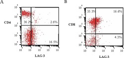
- Expression of LAG-3 on CD4+ and CD8+ subpopulations of tumour infiltrating lymphocytes (TILs) detected with LAG-3 (human), mAb (17B4) (FITC) (Prod. No. ALX-804-806F). Method: TILs from a dissociated renal cell carcinoma sample, stained with 5µg/ml LAG-3 (human), mAb (17B4) (FITC) (Prod. No. ALX-804-806F) and FITC-coupled anti-CD4 or -CD8, are analyzed by a two-colour FACS analysis. Additional staining with anti-CD3 allowed a gate analysis of total T cells. Values indicate percentages in each quadrant [5].

- Submitted by
- Enzo Life Sciences (provider)
- Main image
- Experimental details
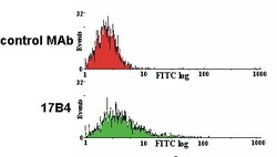
- ÊLAG-3 expression on activated human peripheral blood mononuclear cells (PBMC) detected with LAG-3 (human), mAb (17B4) (FITC)Ê(Prod. No. ALX-804-806F). Method: T lymphocytes from human PBMC are stimulated with 1µg/ml of PHA for three days. Then, after seven days of culture, 3x10E6Êthree-days PHA-activated human PBMC are treated with LAG-3 (human), mAb (17B4) (FITC)Ê(Prod. No. ALX-804-806F) or FITC coupled isotype-matched (IgG1) control MAb (used at a saturating dilution of 1/800 and 1/150 respectively) for 30 min. at 4¡C in RPMI 1640 and washed twice with 1x PBS. Stained cells are then analysed by FACS [4].

- Submitted by
- Enzo Life Sciences (provider)
- Main image
- Experimental details
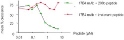
- Specific inhibition of 17B4 staining. Method: LAG-3 (human), mAb (17B4) (FITC)Ê(Prod. No. ALX-804-806F)Ê(10µg/ml) (is preincubated withÊ a specific peptide epitope (208b) or a control tetanus toxoid (TT) peptide at different molarities prior to staining of TILs. Stained cells are then analyzed by FACS.

- Submitted by
- Enzo Life Sciences (provider)
- Main image
- Experimental details
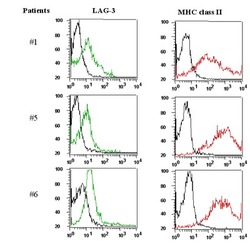
- Tumor infiltrating lymphocytes (TILs) express LAG-3 (detected usingÊLAG-3 (human), mAb (17B4) (Prod. No. ALX-804-806)).ÊMethod: Freshly dissociated single cell suspensions of renal cell carcinoma TILs are incubated withÊLAG-3 (human), mAb (17B4) (FITC)Ê(Prod. No. ALX-804-806) (5µg/ml) and anti-MHC Class II molecules (PE) for 30 min. and washed twice in saline buffer. Additional staining with anti-CD3 allowed a gate analysis of total T cells. The LAG-3 and MHC II profiles of CD3+-gated cells for 3 patients are shown [5].
