 Explore
 Validate
 Learn
Learn Western blot
Antibody data
- Antibody Data
- Antigen structure
- References [1]
- Comments [0]
- Validations
- Western blot [2]
- Immunocytochemistry [1]
Submit
Validation data
Reference
Comment
Report error
- Product number
- MAB8039 - Provider product page

- Provider
- R&D Systems
- Product name
- Human HOIP/RNF31 Antibody
- Antibody type
- Monoclonal
- Description
- Protein A or G purified from hybridoma culture supernatant. Detects human HOIP/RNF31 in direct ELISA and Western Blot.
- Reactivity
- Human
- Host
- Mouse
- Conjugate
- Unconjugated
- Antigen sequence
Q96EP0- Isotype
- IgG
- Antibody clone number
- 875227
- Vial size
- 100 ug
- Storage
- Use a manual defrost freezer and avoid repeated freeze-thaw cycles. 12 months from date of receipt, -20 to -70 °C as supplied. 1 month, 2 to 8 °C under sterile conditions after reconstitution. 6 months, -20 to -70 °C under sterile conditions after reconstitution.
Submitted references Ring finger protein 31-mediated atypical ubiquitination stabilizes forkhead box P3 and thereby stimulates regulatory T-cell function.
Zhu F, Yi G, Liu X, Zhu F, Zhao A, Wang A, Zhu R, Chen Z, Zhao B, Fang S, Yu X, Lin R, Liang R, Li D, Zhao W, Zhang Z, Guo W, Zhang S, Ge S, Fan X, Zhao G, Li B
The Journal of biological chemistry 2018 Dec 28;293(52):20099-20111
The Journal of biological chemistry 2018 Dec 28;293(52):20099-20111
No comments: Submit comment
Supportive validation
- Submitted by
- R&D Systems (provider)
- Main image
- Experimental details
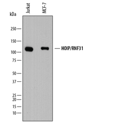
- Detection of Human HOIP/RNF31 by Western Blot. Western blot shows lysates of Jurkat human acute T cell leukemia cell line and MCF-7 human breast cancer cell line. PVDF membrane was probed with 0.5 µg/mL of Mouse Anti-Human HOIP/RNF31 Monoclonal Antibody (Catalog # MAB8039) followed by HRP-conjugated Anti-Mouse IgG Secondary Antibody (Catalog # HAF018). A specific band was detected for HOIP/RNF31 at approximately 120 kDa (as indicated). This experiment was conducted under reducing conditions and using Immunoblot Buffer Group 1.

- Submitted by
- R&D Systems (provider)
- Main image

- Experimental details
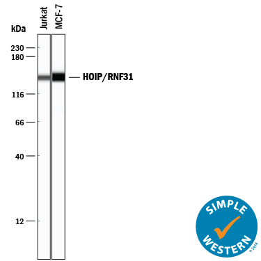
- Detection of Human HOIP/RNF31 by Simple WesternTM. Simple Western lane view shows lysates of Jurkat human acute T cell leukemia cell line and MCF-7 human breast cancer cell line, loaded at 0.5 mg/mL. A specific band was detected for HOIP/RNF31 at approximately 144 kDa (as indicated) using 5 µg/mL of Mouse Anti-Human HOIP/RNF31 Monoclonal Antibody (Catalog # MAB8039). This experiment was conducted under reducing conditions and using the 12-230 kDa separation system.
Supportive validation
- Submitted by
- R&D Systems (provider)
- Main image
- Experimental details
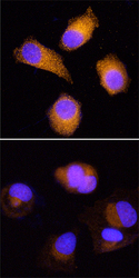
- HOIP/RNF31 in MCF-7 Human Cell Line. HOIP/RNF31 was detected in immersion fixed MCF-7 human breast cancer cell line, untreated (upper panel) or treated with Proteasome Inhibitor I (Tocris Bioscience, Catalog # 4045, lower panel), using Mouse Anti-Human HOIP/RNF31 Monoclonal Antibody (Catalog # MAB8039) at 25 µg/mL for 3 hours at room temperature. Cells were stained using the NorthernLights™ 557-conjugated Anti-Mouse IgG Secondary Antibody (red; Catalog # NL007) and counterstained with DAPI (blue). Specific staining was localized to cytoplasm. View our protocol for Fluorescent ICC Staining of Cells on Coverslips.
