Explore
 Validate
 Learn
Learn Western blot
Antibody data
- Antibody Data
- Antigen structure
- References [1]
- Comments [0]
- Validations
- Western blot [2]
- Immunocytochemistry [2]
- Immunohistochemistry [2]
Submit
Validation data
Reference
Comment
Report error
- Product number
- TA500038 - Provider product page

- Provider
- OriGene
- Proper citation
- OriGene Cat#TA500038, RRID:AB_2162384
- Product name
- Pdx1 mouse monoclonal antibody, clone OTI2A12 (formerly 2A12)
- Antibody type
- Monoclonal
- Description
- Pdx1 mouse monoclonal antibody, clone OTI2A12 (formerly 2A12)
- Host
- Mouse
- Conjugate
- Unconjugated
- Epitope
- PDX1
- Isotype
- IgG
- Antibody clone number
- OTI2A12
- Vial size
- 100 µl
- Concentration
- 0.23 mg/ml
Submitted references Interferon-γ Decreases Nuclear Localization of Pdx-1 and Triggers β-Cell Dysfunction in Chronic Pancreatitis.
Pondugala PK, Sasikala M, Guduru VR, Rebala P, Nageshwar Reddy D
Journal of interferon & cytokine research : the official journal of the International Society for Interferon and Cytokine Research 2015 Jul;35(7):523-9
Journal of interferon & cytokine research : the official journal of the International Society for Interferon and Cytokine Research 2015 Jul;35(7):523-9
No comments: Submit comment
Supportive validation
- Submitted by
- OriGene (provider)
- Main image
- Experimental details
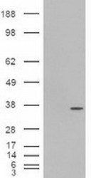
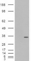
- HEK293T cells were transfected with the pCMV6-ENTRY control (Left lane) or pCMV6-ENTRY PDX1 (RC222354, Right lane) cDNA for 48 hrs and lysed. Equivalent amounts of cell lysates (5 ug per lane) were separated by SDS-PAGE and immunoblotted with anti-PDX1.
- Validation comment
- WB
- Submitted by
- OriGene (provider)
- Main image
- Experimental details
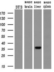
- Western blot analysis of extracts (10ug) from a mouse cell line and 3 different mouse tissues by using anti-PDX1 monoclonal antibody.(1:200)
- Validation comment
- WB

Supportive validation
- Submitted by
- OriGene (provider)
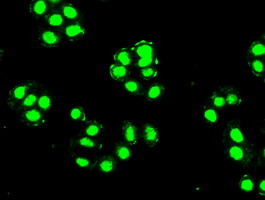
- Main image

- Experimental details
- Immunofluorescent staining of HT29 cells using anti-PDX1 mouse monoclonal antibody (TA500038).
- Validation comment
- IF
- Submitted by
- OriGene (provider)
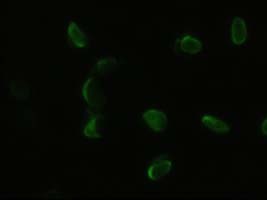
- Main image

- Experimental details
- Anti-PDX1 mouse monoclonal antibody (TA500038) immunofluorescent staining of HELA cells transiently transfected by pCMV6-ENTRY PDX1(RC222354).
- Validation comment
- IF
Supportive validation
- Submitted by
- OriGene (provider)
- Main image
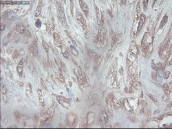
- Experimental details
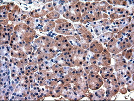
- Immunohistochemical staining of paraffin-embedded Carcinoma of Human pancreas tissue using anti-PDX1 mouse monoclonal antibody. (Heat-induced epitope retrieval by 10mM citric buffer, pH6.0, 100C for 10min, TA500038)
- Validation comment
- IHC
- Submitted by
- OriGene (provider)
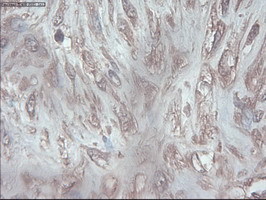
- Main image

- Experimental details
- Immunohistochemical staining of paraffin-embedded Human pancreas tissue within the normal limits using anti-PDX1 mouse monoclonal antibody. (Heat-induced epitope retrieval by 10mM citric buffer, pH6.0, 100C for 10min, TA500038)
- Validation comment
- IHC