 Explore
 Validate
 Learn
Learn Western blot
Antibody data
- Antibody Data
- Antigen structure
- References [1]
- Comments [0]
- Validations
- Western blot [6]
- Immunohistochemistry [2]
Submit
Validation data
Reference
Comment
Report error
- Product number
- PA5-78024 - Provider product page

- Provider
- Invitrogen Antibodies
- Product name
- PDX1 Polyclonal Antibody
- Antibody type
- Polyclonal
- Antigen
- Synthetic peptide
- Description
- Positive Control: Jurkat, Raji, NCI-H929, mouse liver, rat liver, AsPC-1, mouse pancreas Predicted Reactivity: Dog (100%), Pig (100%) Store product as a concentrated solution. Centrifuge briefly prior to opening the vial.
- Reactivity
- Human, Mouse, Rat
- Host
- Rabbit
- Isotype
- IgG
- Vial size
- 100 µL
- Concentration
- 1.29 mg/mL
- Storage
- Store at 4°C short term. For long term storage, store at -20°C, avoiding freeze/thaw cycles.
Submitted references Identification of novel, clinically correlated autoantigens in the monogenic autoimmune syndrome APS1 by proteome-wide PhIP-Seq.
Vazquez SE, Ferré EM, Scheel DW, Sunshine S, Miao B, Mandel-Brehm C, Quandt Z, Chan AY, Cheng M, German M, Lionakis M, DeRisi JL, Anderson MS
eLife 2020 May 15;9
eLife 2020 May 15;9
No comments: Submit comment
Supportive validation
- Submitted by
- Invitrogen Antibodies (provider)
- Main image
- Experimental details
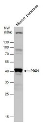
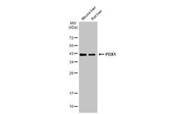
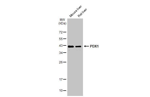
- Western Blot using PDX1 Polyclonal Antibody (Product # PA5-78024). Mouse tissue extract (50 µg) was separated by 12% SDS-PAGE, and the membrane was blotted with PDX1 Polyclonal Antibody (Product # PA5-78024) diluted at 1:1,000. The HRP-conjugated anti-rabbit IgG antibody was used to detect the primary antibody.
- Submitted by
- Invitrogen Antibodies (provider)
- Main image
- Experimental details
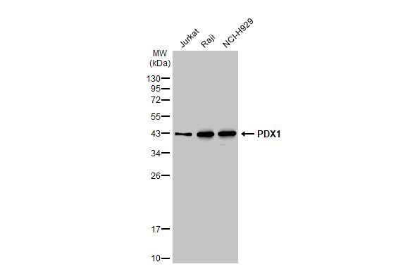
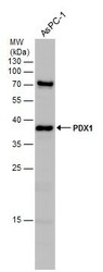
- Western Blot using PDX1 Polyclonal Antibody (Product # PA5-78024). Various whole cell extracts (30 µg) were separated by 12% SDS-PAGE, and the membrane was blotted with PDX1 Polyclonal Antibody (Product # PA5-78024) diluted at 1:1,000. The HRP-conjugated anti-rabbit IgG antibody was used to detect the primary antibody.
- Submitted by
- Invitrogen Antibodies (provider)
- Main image
- Experimental details
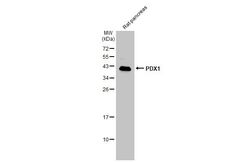
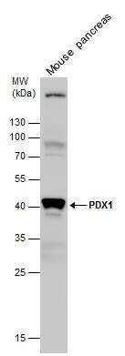
- PDX1 Polyclonal Antibody detects PDX1 protein by western blot analysis. Mouse tissue extracts (50 µg) was separated by 12 % SDS-PAGE, and the membrane was blotted with PDX1 Polyclonal Antibody (Product # PA5-78024) at a dilution of 1:1,000.
- Submitted by
- Invitrogen Antibodies (provider)
- Main image
- Experimental details
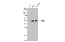
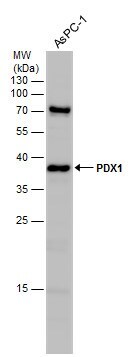
- PDX1 Polyclonal Antibody detects PDX1 protein by western blot analysis. Whole cell extracts (30 µg) was separated by 12% SDS-PAGE, and the membrane was blotted with PDX1 Polyclonal Antibody (Product # PA5-78024) at a dilution of 1:1,000.
- Submitted by
- Invitrogen Antibodies (provider)
- Main image
- Experimental details
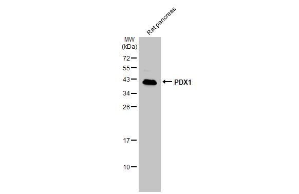
- Western Blot using PDX1 Polyclonal Antibody (Product # PA5-78024). Various tissue extracts (50 µg) were separated by 12% SDS-PAGE, and the membrane was blotted with PDX1 Polyclonal Antibody (Product # PA5-78024) diluted at 1:1,000. The HRP-conjugated anti-rabbit IgG antibody was used to detect the primary antibody.
- Submitted by
- Invitrogen Antibodies (provider)
- Main image
- Experimental details
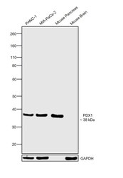
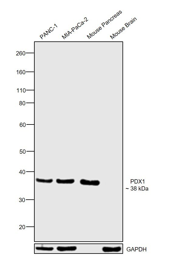
- Western blot was performed using Anti-PDX1 Polyclonal Antibody (Product # PA5-78024) and a 38 kDa band corresponding to PDX1 was observed across cell lines and tissues tested except Mouse Brain which is reported to be negative. Whole cell extracts (30 µg lysate) of PANC-1 (Lane 1), MIA-PaCa-2 (Lane 2), tissue extracts of Mouse Pancreas (Lane 3) and Mouse Brain (Lane 4) were electrophoresed using NuPAGE™ 4-12% Bis-Tris Protein Gel (Product # NP0322BOX). Resolved proteins were then transferred onto a nitrocellulose membrane (Product # IB23001) by iBlot® 2 Dry Blotting System (Product # IB21001). The blot was probed with the primary antibody (1:3000 dilution) and detected by chemiluminescence with Goat anti-Rabbit IgG (Heavy Chain) Superclonal™ Recombinant Secondary Antibody, HRP (Product # A27036, 1:4000 dilution) using the iBright FL 1000 (Product # A32752). Chemiluminescent detection was performed using Novex® ECL Chemiluminescent Substrate Reagent Kit (Product # WP20005).
Supportive validation
- Submitted by
- Invitrogen Antibodies (provider)
- Main image
- Experimental details
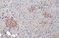
- PDX1 Polyclonal Antibody detects PDX1 protein at nucleus by immunohistochemical analysis. Sample: Paraffin-embedded mouse pancreas. PDX1 stained by PDX1 Polyclonal Antibody (Product # PA5-78024) diluted at 1:500. Antigen Retrieval: Citrate buffer, pH 6.0, 15 min.

- Submitted by
- Invitrogen Antibodies (provider)
- Main image
- Experimental details
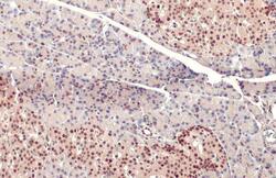
- PDX1 Polyclonal Antibody detects PDX1 protein at nucleus by immunohistochemical analysis. Sample: Paraffin-embedded rat pancreas. PDX1 stained by PDX1 Polyclonal Antibody (Product # PA5-78024) diluted at 1:500. Antigen Retrieval: Citrate buffer, pH 6.0, 15 min.
