 Explore
 Validate
 Learn
Learn Western blot
 Immunohistochemistry
Antibody data
- Antibody Data
- Antigen structure
- References [5]
- Comments [0]
- Validations
- Western blot [1]
- Immunocytochemistry [2]
Submit
Validation data
Reference
Comment
Report error
- Product number
- MAB2419 - Provider product page

- Provider
- R&D Systems
- Product name
- Human/Mouse PDX-1/IPF1 Antibody
- Antibody type
- Monoclonal
- Description
- Protein A or G purified from hybridoma culture supernatant. Detects human and mouse PDX-1 in Western blots.
- Reactivity
- Human, Mouse
- Host
- Mouse
- Conjugate
- Unconjugated
- Antigen sequence
P52945- Isotype
- IgG
- Antibody clone number
- 267712
- Vial size
- 100 ug
- Concentration
- LYOPH
- Storage
- Use a manual defrost freezer and avoid repeated freeze-thaw cycles. 12 months from date of receipt, -20 to -70 °C as supplied. 1 month, 2 to 8 °C under sterile conditions after reconstitution. 6 months, -20 to -70 °C under sterile conditions after reconstitution.
Submitted references A programmable synthetic lineage-control network that differentiates human IPSCs into glucose-sensitive insulin-secreting beta-like cells.
Redifferentiation of adult human β cells expanded in vitro by inhibition of the WNT pathway.
Peroxisome proliferator-activated receptor gamma activation restores islet function in diabetic mice through reduction of endoplasmic reticulum stress and maintenance of euchromatin structure.
Betacellulin and nicotinamide sustain PDX1 expression and induce pancreatic beta-cell differentiation in human embryonic stem cells.
Generation of insulin-producing cells from PDX-1 gene-modified human mesenchymal stem cells.
Saxena P, Heng BC, Bai P, Folcher M, Zulewski H, Fussenegger M
Nature communications 2016 Apr 11;7:11247
Nature communications 2016 Apr 11;7:11247
Redifferentiation of adult human β cells expanded in vitro by inhibition of the WNT pathway.
Lenz A, Toren-Haritan G, Efrat S
PloS one 2014;9(11):e112914
PloS one 2014;9(11):e112914
Peroxisome proliferator-activated receptor gamma activation restores islet function in diabetic mice through reduction of endoplasmic reticulum stress and maintenance of euchromatin structure.
Evans-Molina C, Robbins RD, Kono T, Tersey SA, Vestermark GL, Nunemaker CS, Garmey JC, Deering TG, Keller SR, Maier B, Mirmira RG
Molecular and cellular biology 2009 Apr;29(8):2053-67
Molecular and cellular biology 2009 Apr;29(8):2053-67
Betacellulin and nicotinamide sustain PDX1 expression and induce pancreatic beta-cell differentiation in human embryonic stem cells.
Cho YM, Lim JM, Yoo DH, Kim JH, Chung SS, Park SG, Kim TH, Oh SK, Choi YM, Moon SY, Park KS, Lee HK
Biochemical and biophysical research communications 2008 Feb 1;366(1):129-34
Biochemical and biophysical research communications 2008 Feb 1;366(1):129-34
Generation of insulin-producing cells from PDX-1 gene-modified human mesenchymal stem cells.
Li Y, Zhang R, Qiao H, Zhang H, Wang Y, Yuan H, Liu Q, Liu D, Chen L, Pei X
Journal of cellular physiology 2007 Apr;211(1):36-44
Journal of cellular physiology 2007 Apr;211(1):36-44
No comments: Submit comment
Supportive validation
- Submitted by
- R&D Systems (provider)
- Main image

- Experimental details
- Detection of Mouse PDX-1/IPF1 by Western Blot. Western blot shows lysates of beta TC-6 mouse beta cell insulinoma cell line. PVDF membrane was probed with 1 µg/mL of Mouse Anti-Human/Mouse PDX-1/IPF1 Monoclonal Antibody (Catalog # MAB2419) followed by HRP-conjugated Anti-Mouse IgG Secondary Antibody (Catalog # HAF007). A specific band was detected for PDX-1/IPF1 at approximately 46 kDa (as indicated). This experiment was conducted under reducing conditions and using Immunoblot Buffer Group 1.

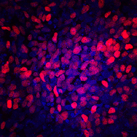
Supportive validation
- Submitted by
- R&D Systems (provider)
- Main image

- Experimental details
- PDX-1/IPF1 in BG01V Human Embryonic Stem Cells. PDX-1/IPF1 was detected in immersion fixed BG01V human embryonic stem cells differentiated into pancreatic progenitor cells using Mouse Anti-Human/Mouse PDX-1/IPF1 Monoclonal Antibody (Catalog # MAB2419) at 10 µg/mL for 3 hours at room temperature. Cells were stained using the NorthernLights™ 557-conjugated Anti-Mouse IgG Secondary Antibody (red; Catalog # NL007) and counterstained with DAPI (blue). Specific staining was localized to nuclei. View our protocol for Fluorescent ICC Staining of Stem Cells on Coverslips.
- Submitted by
- R&D Systems (provider)
- Main image

- Experimental details
- PDX-1/IPF1 in beta TC-6 Mouse Cell Line. PDX-1/IPF1 was detected in immersion fixed beta TC-6 mouse beta cell insulinoma cell line using Human/Mouse PDX-1/IPF1 Monoclonal Antibody (Catalog # MAB2419) at 10 µg/mL for 3 hours at room temperature. Cells were stained using the NorthernLights™ 557-conjugated Anti-Mouse IgG Secondary Antibody (yellow, upper panel; Catalog # NL007) and counterstained with DAPI (blue, lower panel). View our protocol for Fluorescent ICC Staining of Cells on Coverslips.
