 Explore
 Validate
 Learn
LearnPA5-17436
antibody from Invitrogen Antibodies
Targeting: PNPLA2
ATGL, desnutrin, FP17548, iPLA2zeta, TTS-2.2
 Western blot
 Immunocytochemistry
 Immunoprecipitation
Antibody data
- Antibody Data
- Antigen structure
- References [1]
- Comments [0]
- Validations
- Immunocytochemistry [5]
Submit
Validation data
Reference
Comment
Report error
- Product number
- PA5-17436 - Provider product page

- Provider
- Invitrogen Antibodies
- Product name
- ATGL Polyclonal Antibody
- Antibody type
- Polyclonal
- Antigen
- Synthetic peptide
- Description
- It is not recommended to aliquot this antibody.
- Reactivity
- Human, Mouse
- Host
- Rabbit
- Isotype
- IgG
- Vial size
- 100 μL
- Concentration
- 129 μg/mL
- Storage
- -20°C
Submitted references Carboxyl Ester Lipase May Not Mediate Lipotoxic Injury during Severe Acute Pancreatitis.
Khatua B, Trivedi RN, Noel P, Patel K, Singh R, de Oliveira C, Trivedi S, Mishra V, Lowe M, Singh VP
The American journal of pathology 2019 Jun;189(6):1226-1240
The American journal of pathology 2019 Jun;189(6):1226-1240
No comments: Submit comment
Supportive validation
- Submitted by
- Invitrogen Antibodies (provider)
- Main image

- Experimental details
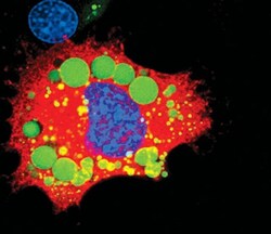
- Immunofluorescent analysis of ATGL in 3T3-L1 cells using an ATGL polyclonal antibody (Product # PA5-17436) (red) showing cytoplasmic localization in differentiated cells. Lipid droplets have been labeled with a BODIPY fluorescent dye (green). DNA is labeled using a fluorescent blue dye.

- Submitted by
- Invitrogen Antibodies (provider)
- Main image
- Experimental details
- Immunofluorescent analysis of ATGL in 3T3-L1 cells using an ATGL polyclonal antibody (Product # PA5-17436) (red) showing cytoplasmic localization in differentiated cells. Lipid droplets have been labeled with a BODIPY fluorescent dye (green). DNA is labeled using a fluorescent blue dye.

- Submitted by
- Invitrogen Antibodies (provider)
- Main image

- Experimental details
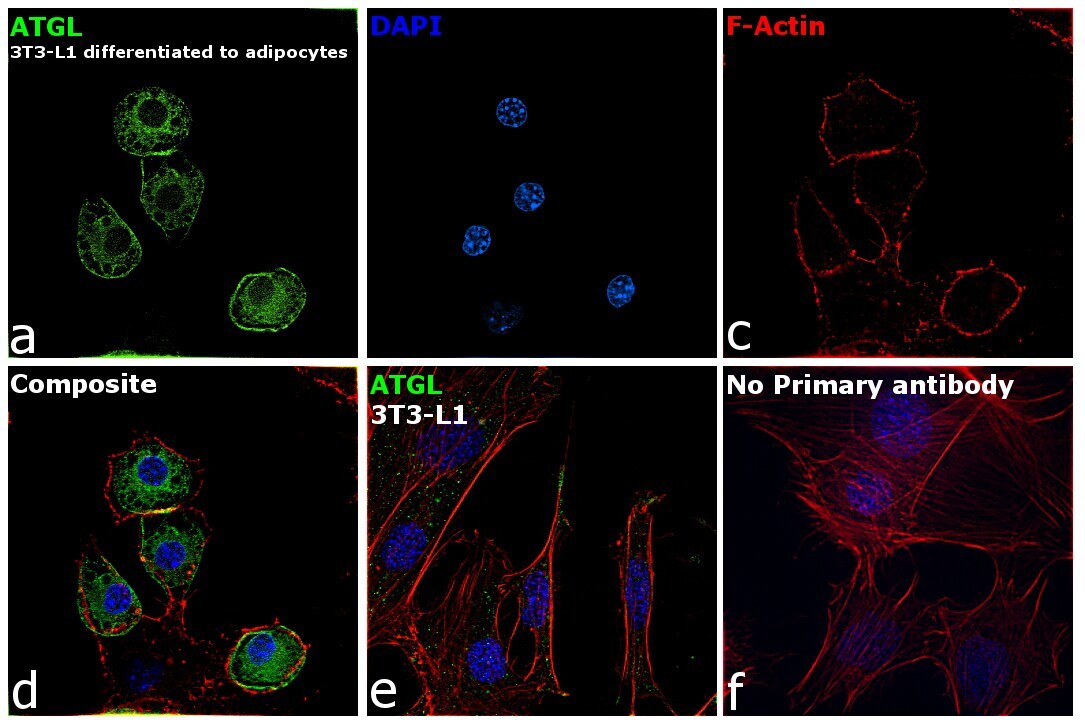
- Immunofluorescence analysis of ATGL was performed using 70% confluent log phase 3T3-L1 and 3T3-L1 differentiated to adipocytes. The cells were fixed with 4% paraformaldehyde for 10 minutes, permeabilized with 0.1% Triton™ X-100 for 15 minutes, and blocked with 2% BSA for 1 hour at room temperature. The cells were labeled with ATGL Polyclonal Antibody (Product # PA5-17436) at 1:100 dilution in 0.1% BSA, incubated at 4 degree Celsius overnight and then labeled with Goat anti-Rabbit IgG (H+L) Superclonal™ Recombinant Secondary Antibody, Alexa Fluor® 488 conjugate (Product # A27034) at a dilution of 1:2000 for 45 minutes at room temperature (Panel a: green). Nuclei (Panel b: blue) were stained with SlowFade® Gold Antifade Mountant with DAPI (Product # S36938). F-actin (Panel c: red) was stained with Rhodamine Phalloidin (Product # R415, 1:300). Panel d represents the merged image showing localization to vacuolar structure (fat droplets) in cytoplasm. Panel e shows 3T3-L1 cells with less expression of ATGL. Panel f represents control cells with no primary antibody to assess background. The images were captured at 60X magnification.
- Submitted by
- Invitrogen Antibodies (provider)
- Main image

- Experimental details
- Immunofluorescent analysis of ATGL in 3T3-L1 cells using an ATGL polyclonal antibody (Product # PA5-17436) (red) showing cytoplasmic localization in differentiated cells. Lipid droplets have been labeled with a BODIPY fluorescent dye (green). DNA is labeled using a fluorescent blue dye.

- Submitted by
- Invitrogen Antibodies (provider)
- Main image
- Experimental details
- Immunofluorescence analysis of ATGL was performed using 70% confluent log phase 3T3-L1 and 3T3-L1 differentiated to adipocytes. The cells were fixed with 4% paraformaldehyde for 10 minutes, permeabilized with 0.1% Triton™ X-100 for 15 minutes, and blocked with 2% BSA for 1 hour at room temperature. The cells were labeled with ATGL Polyclonal Antibody (Product # PA5-17436) at 1:100 dilution in 0.1% BSA, incubated at 4 degree Celsius overnight and then labeled with Goat anti-Rabbit IgG (Heavy Chain) Superclonal™ Recombinant Secondary Antibody, Alexa Fluor® 488 conjugate (Product # A27034) at a dilution of 1:2000 for 45 minutes at room temperature (Panel a: green). Nuclei (Panel b: blue) were stained with SlowFade® Gold Antifade Mountant with DAPI (Product # S36938). F-actin (Panel c: red) was stained with Rhodamine Phalloidin (Product # R415, 1:300). Panel d represents the merged image showing localization to vacuolar structure (fat droplets) in cytoplasm. Panel e shows 3T3-L1 cells with less expression of ATGL. Panel f represents control cells with no primary antibody to assess background. The images were captured at 60X magnification.
