Explore
 Validate
 Learn
Learn Western blot
 ELISA
ELISA Immunocytochemistry
Antibody data
- Antibody Data
- Antigen structure
- References [4]
- Comments [0]
- Validations
- Western blot [10]
- Immunocytochemistry [2]
- Immunohistochemistry [1]
Submit
Validation data
Reference
Comment
Report error
- Product number
- GTX104618 - Provider product page

- Provider
- GeneTex
- Proper citation
- GeneTex Cat#GTX104618, RRID:AB_1241405
- Product name
- Triosephosphate isomerase antibody [C2C3], C-term
- Antibody type
- Polyclonal
- Reactivity
- Human, Mouse, Rat
- Host
- Rabbit
Submitted references Proteomic profiling identifies outcome-predictive markers in patients with peripheral T-cell lymphoma, not otherwise specified.
High glucose-induced proteome alterations in hepatocytes and its possible relevance to diabetic liver disease.
A Study of the Wound Healing Mechanism of a Traditional Chinese Medicine, Angelica sinensis, Using a Proteomic Approach.
Differential expression of fourteen proteins between uveal melanoma from patients who subsequently developed distant metastases versus those who did Not.
Ludvigsen M, Bjerregård Pedersen M, Lystlund Lauridsen K, Svenstrup Poulsen T, Hamilton-Dutoit SJ, Besenbacher S, Bendix K, Møller MB, Nørgaard P, d'Amore F, Honoré B
Blood advances 2018 Oct 9;2(19):2533-2542
Blood advances 2018 Oct 9;2(19):2533-2542
High glucose-induced proteome alterations in hepatocytes and its possible relevance to diabetic liver disease.
Chen JY, Chou HC, Chen YH, Chan HL
The Journal of nutritional biochemistry 2013 Nov;24(11):1889-910
The Journal of nutritional biochemistry 2013 Nov;24(11):1889-910
A Study of the Wound Healing Mechanism of a Traditional Chinese Medicine, Angelica sinensis, Using a Proteomic Approach.
Hsiao CY, Hung CY, Tsai TH, Chak KF
Evidence-based complementary and alternative medicine : eCAM 2012;2012:467531
Evidence-based complementary and alternative medicine : eCAM 2012;2012:467531
Differential expression of fourteen proteins between uveal melanoma from patients who subsequently developed distant metastases versus those who did Not.
Linge A, Kennedy S, O'Flynn D, Beatty S, Moriarty P, Henry M, Clynes M, Larkin A, Meleady P
Investigative ophthalmology & visual science 2012 Jul 9;53(8):4634-43
Investigative ophthalmology & visual science 2012 Jul 9;53(8):4634-43
No comments: Submit comment
Enhanced validation
Supportive validation
- Submitted by
- GeneTex (provider)
- Enhanced method
- Genetic validation
- Main image
- Experimental details
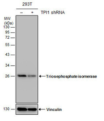
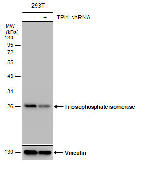
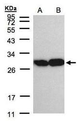
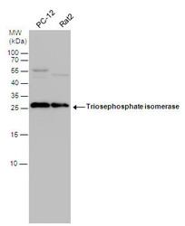
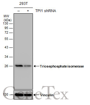
- Non-transfected (¡V) and transfected (+) 293T whole cell extracts (30 ?g) were separated by 12% SDS-PAGE, and the membrane was blotted with Triosephosphate isomerase antibody [C2C3], C-term (GTX104618) diluted at 1:3000. The HRP-conjugated anti-rabbit IgG antibody (GTX213110-01) was used to detect the primary antibody.
Supportive validation
- Submitted by
- GeneTex (provider)
- Main image

- Experimental details
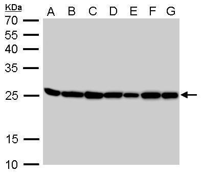
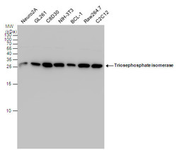
- Triosephosphate isomerase antibody [C2C3], C-term detects TPI1 protein by Western blot analysis.A. 30 µg Neuro2A whole cell lysate/extract B. 30 µg GL261 whole cell lysate/extract C. 30 µg C8D30 whole cell lysate/extract D. 30 µg NIH-3T3 whole cell lysate/extract E. 30 µg BCL-1 whole cell lysate/extract F. 30 µg Raw264.7 whole cell lysate/extract G. 30 µg C2C12 whole cell lysate/extract 12 % SDS-PAGETriosephosphate isomerase antibody [C2C3], C-term (GTX104618) dilution: 1:1000
- Validation comment
- WB
- Submitted by
- GeneTex (provider)
- Main image
- Experimental details
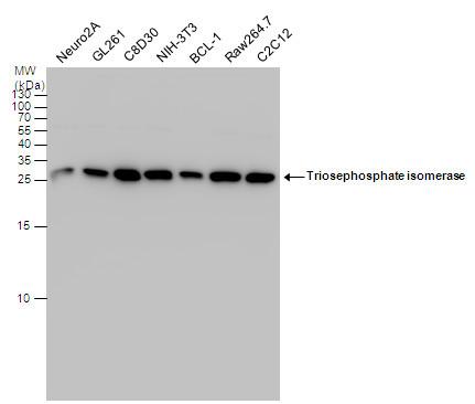
- Triosephosphate isomerase antibody [C2C3], C-term detects TPI1 protein by Western blot analysis.A. 30 µg PC-12 whole cell lysate/extract B. 30 µg Rat2 whole cell lysate/extract12 % SDS-PAGETriosephosphate isomerase antibody [C2C3], C-term (GTX104618) dilution: 1:1000
- Validation comment
- WB
- Submitted by
- GeneTex (provider)
- Main image
- Experimental details
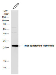
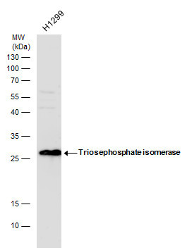
- Sample(30 ?g of whole cell lysate)A:A431(GTX27909)B:H129912% SDS PAGEGTX104618 diluted at 1:2000
- Validation comment
- WB
- Submitted by
- GeneTex (provider)
- Main image
- Experimental details
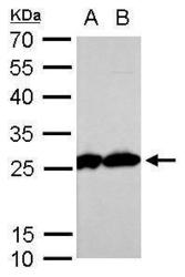
- Triosephosphate isomerase antibody detects Triosephosphate isomerase protein by western blot analysis. Whole cell extracts (30 ?g) was separated by 15% SDS-PAGE, and blotted with Triosephosphate isomerase antibody (GTX104618) diluted by 1:1000. The HRP-conjugated anti-rabbit IgG antibody (GTX213110-01) was used to detect the primary antibody.
- Submitted by
- GeneTex (provider)
- Main image
- Experimental details
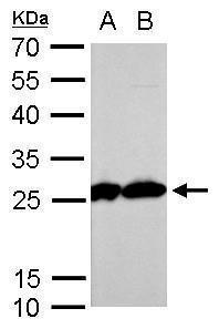
- Triosephosphate isomerase antibody detects Triosephosphate isomerase protein by western blot analysis. Various whole cell extracts (30 ?g) were separated by 15% SDS-PAGE, and the membrane was blotted with Triosephosphate isomerase antibody (GTX104618) diluted by 1:1000. The HRP-conjugated anti-rabbit IgG antibody (GTX213110-01) was used to detect the primary antibody.
- Submitted by
- GeneTex (provider)
- Main image
- Experimental details
- Triosephosphate isomerase antibody detects Triosephosphate isomerase protein by western blot analysis. Various whole cell extracts (30 ?g) were separated by 15% SDS-PAGE, and the membrane was blotted with Triosephosphate isomerase antibody (GTX104618) diluted by 1:1000. The HRP-conjugated anti-rabbit IgG antibody (GTX213110-01) was used to detect the primary antibody.

- Submitted by
- GeneTex (provider)
- Main image
- Experimental details
- Non-transfected (¡V) and transfected (+) 293T whole cell extracts (30 ?g) were separated by 12% SDS-PAGE, and the membrane was blotted with Triosephosphate isomerase antibody [C2C3], C-term (GTX104618) diluted at 1:3000. The HRP-conjugated anti-rabbit IgG antibody (GTX213110-01) was used to detect the primary antibody.
- Submitted by
- GeneTex (provider)
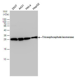
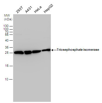
- Main image
- Experimental details
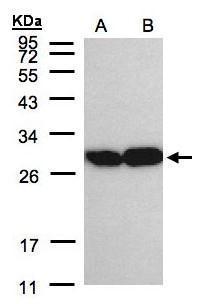
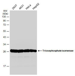
- Various whole cell extracts (30 ?g) were separated by 12% SDS-PAGE, and the membrane was blotted with Triosephosphate isomerase antibody [C2C3], C-term (GTX104618) diluted at 1:1000. The HRP-conjugated anti-rabbit IgG antibody (GTX213110-01) was used to detect the primary antibody.
Supportive validation
- Submitted by
- GeneTex (provider)
- Main image
- Experimental details
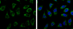
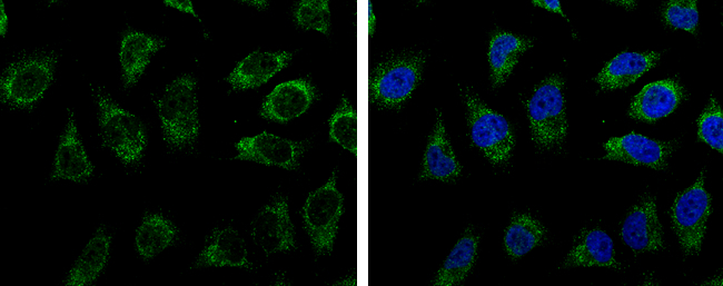
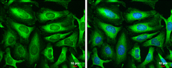
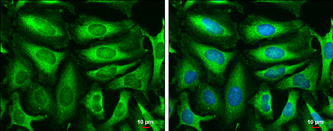
- Triosephosphate isomerase antibody [C2C3], C-term detects Triosephosphate isomerase protein at cytoplasm by immunofluorescent analysis.Sample: HeLa cells were fixed in 4% paraformaldehyde at RT for 15 min.Green: Triosephosphate isomerase protein stained by Triosephosphate isomerase antibody [C2C3], C-term (GTX104618) diluted at 1:500.Blue: Hoechst 33342 staining.Scale bar = 10 £gm.
- Submitted by
- GeneTex (provider)
- Main image
- Experimental details
- Triosephosphate isomerase antibody [C2C3], C-term detects Triosephosphate isomerase protein at cytoplasm by immunofluorescent analysis.Sample: HeLa cells were fixed in 4% paraformaldehyde at RT for 15 min.Green: Triosephosphate isomerase stained by Triosephosphate isomerase antibody [C2C3], C-term (GTX104618) diluted at 1:500.Blue: Hoechst 33342 staining.Scale bar= 10£gm.
Supportive validation
- Submitted by
- GeneTex (provider)
- Main image
- Experimental details
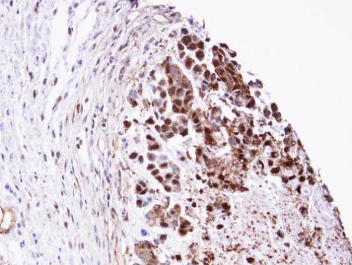
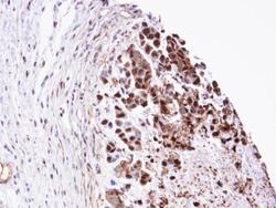
- Immunohistochemical analysis of paraffin-embedded SNU-16 xenograft, using TPI1 (GTX104618) antibody at 1:100 dilution.