Explore
 Validate
 Learn
Learn Western blot
 ELISA
ELISAAntibody data
- Antibody Data
- Antigen structure
- References [7]
- Comments [0]
- Validations
- Western blot [7]
- Immunohistochemistry [1]
Submit
Validation data
Reference
Comment
Report error
- Product number
- NBP1-31470 - Provider product page

- Provider
- Novus Biologicals
- Proper citation
- Novus Cat#NBP1-31470, RRID:AB_2287730
- Product name
- Rabbit Polyclonal Triosephosphate isomerase Antibody
- Antibody type
- Polyclonal
- Description
- Immunogen affinity purified.
- Reactivity
- Human, Mouse, Rat, Bovine
- Host
- Rabbit
- Isotype
- IgG
- Vial size
- 100 ul
- Storage
- Aliquot and store at -20C or -80C. Avoid freeze-thaw cycles.
Submitted references Quantification of biomarkers for beef meat qualities using a combination of Parallel Reaction Monitoring- and antibody-based proteomics.
Beef tenderness and intramuscular fat proteomic biomarkers: Effect of gender and rearing practices.
Beef tenderness and intramuscular fat proteomic biomarkers: muscle type effect.
Reverse Phase Protein array for the quantification and validation of protein biomarkers of beef qualities: The case of meat color from Charolais breed.
Reverse phase protein arrays for the identification/validation of biomarkers of beef texture and their use for early classification of carcasses.
Proteomic analysis of Pteropus alecto kidney cells in response to the viral mimic, Poly I:C.
Externalized glycolytic enzymes are novel, conserved, and early biomarkers of apoptosis.
Bonnet M, Soulat J, Bons J, Léger S, De Koning L, Carapito C, Picard B
Food chemistry 2020 Jul 1;317:126376
Food chemistry 2020 Jul 1;317:126376
Beef tenderness and intramuscular fat proteomic biomarkers: Effect of gender and rearing practices.
Picard B, Gagaoua M, Al Jammas M, Bonnet M
Journal of proteomics 2019 May 30;200:1-10
Journal of proteomics 2019 May 30;200:1-10
Beef tenderness and intramuscular fat proteomic biomarkers: muscle type effect.
Picard B, Gagaoua M, Al-Jammas M, De Koning L, Valais A, Bonnet M
PeerJ 2018;6:e4891
PeerJ 2018;6:e4891
Reverse Phase Protein array for the quantification and validation of protein biomarkers of beef qualities: The case of meat color from Charolais breed.
Gagaoua M, Bonnet M, De Koning L, Picard B
Meat science 2018 Nov;145:308-319
Meat science 2018 Nov;145:308-319
Reverse phase protein arrays for the identification/validation of biomarkers of beef texture and their use for early classification of carcasses.
Gagaoua M, Bonnet M, Ellies-Oury MP, De Koning L, Picard B
Food chemistry 2018 Jun 1;250:245-252
Food chemistry 2018 Jun 1;250:245-252
Proteomic analysis of Pteropus alecto kidney cells in response to the viral mimic, Poly I:C.
Mok L, Wynne JW, Ford K, Shiell B, Bacic A, Michalski WP
Proteome science 2015;13:25
Proteome science 2015;13:25
Externalized glycolytic enzymes are novel, conserved, and early biomarkers of apoptosis.
Ucker DS, Jain MR, Pattabiraman G, Palasiewicz K, Birge RB, Li H
The Journal of biological chemistry 2012 Mar 23;287(13):10325-43
The Journal of biological chemistry 2012 Mar 23;287(13):10325-43
No comments: Submit comment
Supportive validation
- Submitted by
- Novus Biologicals (provider)
- Main image
- Experimental details
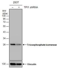
- Western Blot: Triosephosphate isomerase Antibody [NBP1-31470] - Non-transfected (-) and transfected (+) 293T whole cell extracts (30 ug) were separated by 12% SDS-PAGE, and the membrane was blotted with Triosephosphate isomerase antibody [C2C3], C-term. The HRP-conjugated anti-rabbit IgG antibody was used to detect the primary antibody.
- Submitted by
- Novus Biologicals (provider)
- Main image
- Experimental details
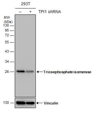
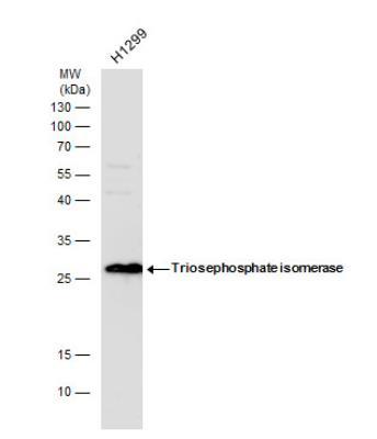
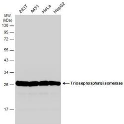
- Western Blot: Triosephosphate isomerase Antibody [NBP1-31470] - Whole cell extracts (30 ug) was separated by 15% SDS-PAGE, and blotted with Triosephosphate isomerase antibody diluted by 1:1000. The HRP-conjugated anti-rabbit IgG antibody (NBP2-19301) was used to detect the primary antibody.
- Submitted by
- Novus Biologicals (provider)
- Main image
- Experimental details
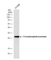
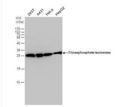
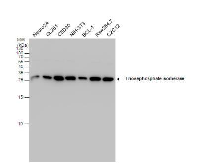
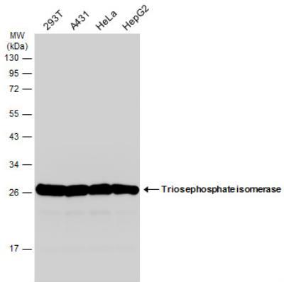
- Western Blot: Triosephosphate isomerase Antibody [NBP1-31470] - Various whole cell extracts (30 ug) were separated by 15% SDS-PAGE, and the membrane was blotted with Triosephosphate isomerase antibody diluted by 1:1000. The HRP-conjugated anti-rabbit IgG antibody (NBP2-19301) was used to detect the primary antibody.
- Submitted by
- Novus Biologicals (provider)
- Main image
- Experimental details
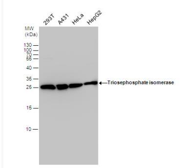
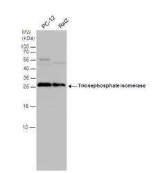
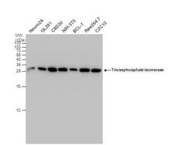
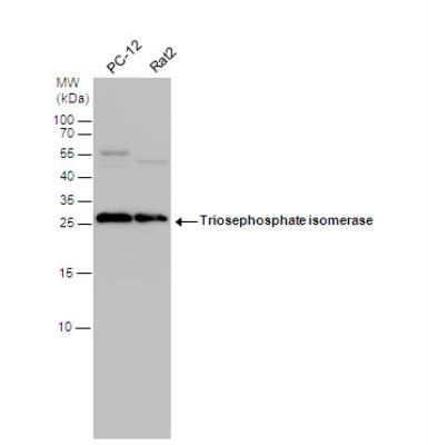
- Western Blot: Triosephosphate isomerase Antibody [NBP1-31470] - Various whole cell extracts (30 ug) were separated by 12% SDS-PAGE, and the membrane was blotted with Triosephosphate isomerase antibody [C2C3], C-term diluted at 1:1000. The HRP-conjugated anti-rabbit IgG antibody (NBP2-19301) was used to detect the primary antibody.
- Submitted by
- Novus Biologicals (provider)
- Main image

- Experimental details
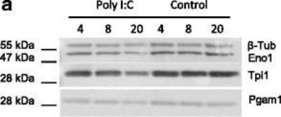
- Western Blot: Triosephosphate isomerase Antibody [NBP1-31470] - Immunodetection of glycolytic enzymes within bat and human cells. Immunodetection of Eno1, Tpi1, Pgam1 and beta2-Tub (load control) in Poly I:C transfected and Control PaKiT03 cells at 4, 8 and 20 hpt. Image collected and cropped by CiteAb from the following publication (http://www.proteomesci.com/content/13/1/25), licensed under a CC-BY licence.
Supportive validation
- Submitted by
- Novus Biologicals (provider)
- Main image
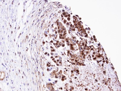
- Experimental details
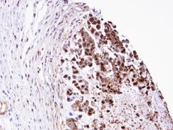
- Immunohistochemistry-Paraffin: Triosephosphate isomerase Antibody [NBP1-31470] - Paraffin-embedded SNU-16 xenograft, using antibody at 1:100 dilution.