Explore
 Validate
 Learn
Learn Western blot
 Immunocytochemistry
 Immunoprecipitation
Antibody data
- Antibody Data
- Antigen structure
- References [2]
- Comments [0]
- Validations
- Immunocytochemistry [1]
- Immunohistochemistry [1]
- Other assay [1]
Submit
Validation data
Reference
Comment
Report error
- Product number
- PA5-17476 - Provider product page

- Provider
- Invitrogen Antibodies
- Product name
- NEDD8 Polyclonal Antibody
- Antibody type
- Polyclonal
- Antigen
- Synthetic peptide
- Description
- It is not recommended to aliquot this antibody. This antibody is not cross-reactive with other ubiquitin family members, including ubiquitin, SUMO1, SUMO2, SUMO3 and ISG15.
- Reactivity
- Human, Mouse, Rat
- Host
- Rabbit
- Isotype
- IgG
- Vial size
- 100 μL
- Concentration
- 162 μg/mL
- Storage
- -20°C
Submitted references Effects of the NEDD8-Activating Enzyme Inhibitor MLN4924 on Lytic Reactivation of Kaposi's Sarcoma-Associated Herpesvirus.
Effects of Exogenous NUB1 Expression in the Striatum of HDQ175/Q7 Mice.
Chang PJ, Chen LW, Chen LY, Hung CH, Shih YJ, Wang SS
Journal of virology 2017 Oct 1;91(19)
Journal of virology 2017 Oct 1;91(19)
Effects of Exogenous NUB1 Expression in the Striatum of HDQ175/Q7 Mice.
Vodicka P, Chase K, Iuliano M, Valentine DT, Sapp E, Lu B, Kegel-Gleason KB, Sena-Esteves M, Aronin N, DiFiglia M
Journal of Huntington's disease 2016 Jun 13;5(2):163-74
Journal of Huntington's disease 2016 Jun 13;5(2):163-74
No comments: Submit comment
Supportive validation
- Submitted by
- Invitrogen Antibodies (provider)
- Main image

- Experimental details
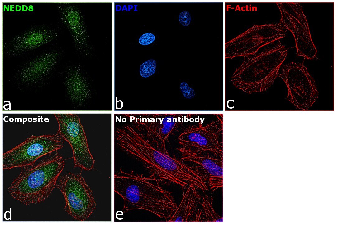
- Immunofluorescence analysis of NEDD8 was performed using 70% confluent log phase HeLa cells. The cells were fixed with 4% paraformaldehyde for 10 minutes, permeabilized with 0.1% Triton™ X-100 for 15 minutes, and blocked with 2% BSA for 1 hour at room temperature. The cells were labeled with NEDD8 Polyclonal Antibody (Product # PA5-17476) at 1:100 dilution in 0.1% BSA, incubated at 4 degree Celsius overnight and then labeled with Goat anti-Rabbit IgG (Heavy Chain) Superclonal™ Recombinant Secondary Antibody, Alexa Fluor® 488 conjugate (Product # A27034) at a dilution of 1:2000 for 45 minutes at room temperature (Panel a: green). Nuclei (Panel b: blue) were stained with SlowFade® Gold Antifade Mountant with DAPI (Product # S36938). F-actin (Panel c: red) was stained with Rhodamine Phalloidin (Product # R415, 1:300). Panel d represents the merged image showing localization to the cytoplasm and nucleus. Panel e represents control cells with no primary antibody to assess background. The images were captured at 60X magnification.
Supportive validation
- Submitted by
- Invitrogen Antibodies (provider)
- Main image
- Experimental details
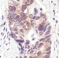
- Immunohistochemical analysis of NEDD8 in paraffin-embedded human colon carcinoma using a NEDD8 polyclonal antibody (Product # PA5-17476) showing cytoplasmic and membrane localization.

Supportive validation
- Submitted by
- Invitrogen Antibodies (provider)
- Main image

- Experimental details
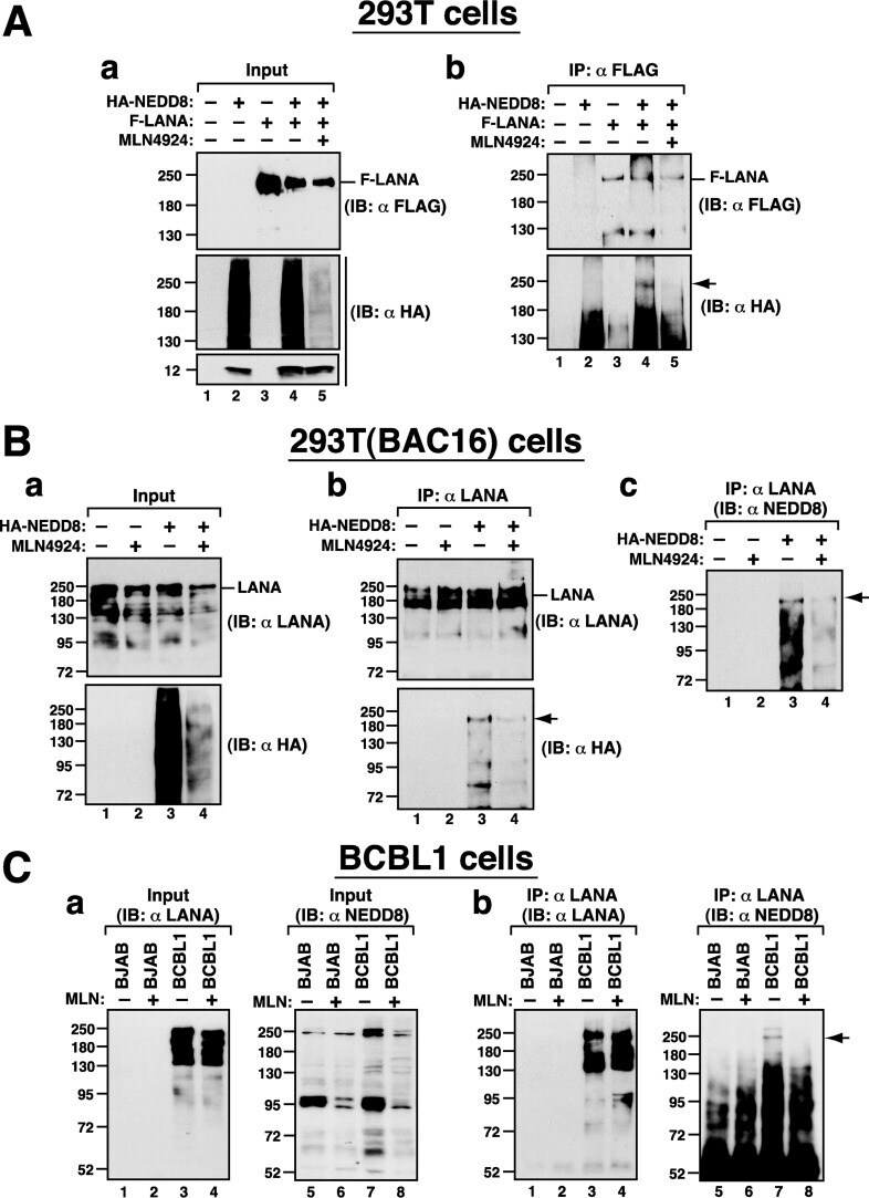
- FIG 6 LANA is naturally modified with NEDD8 in cells. (A) Neddylation of LANA in 293T cells. 293T cells were transfected with the indicated plasmids expressing HA-NEDD8 or F-LANA and left untreated or treated with MLN4924 (2.0 muM) for 24 h. Cell samples were then subjected to immunoprecipitation (IP) and immunoblot (IB) analysis. (B) Neddylation of LANA in 293T(BAC16) cells. 293T(BAC16) cells were transfected with the expression plasmid for HA-NEDD8 and cultured in medium with or without MLN4924 (2.0 muM) for 24 h. Cell lysates were immunoprecipitated using anti-LANA antibody, and the resultant immunoprecipitates were analyzed by immunoblotting using antibodies against LANA, HA, and NEDD8. (C) Neddylation of LANA in BCBL1 cells. BJAB and BCBL1 cells were left untreated or treated with MLN4924 (2.0 muM) for 24 h. After cell lysates were immunoprecipitated with anti-LANA antibody, the immunoprecipitated proteins were probed with anti-LANA and anti-NEDD8 antibodies. Arrows indicate the positions of the neddylated LANA.