Explore
 Validate
 Learn
Learn Western blot
 Immunocytochemistry
Antibody data
- Antibody Data
- Antigen structure
- References [1]
- Comments [0]
- Validations
- Immunocytochemistry [1]
- Other assay [1]
Submit
Validation data
Reference
Comment
Report error
- Product number
- 702694 - Provider product page

- Provider
- Invitrogen Antibodies
- Product name
- LOXL2 Recombinant Rabbit Monoclonal Antibody (7H25L21)
- Antibody type
- Monoclonal
- Antigen
- Synthetic peptide
- Reactivity
- Human
- Host
- Rabbit
- Isotype
- IgG
- Antibody clone number
- 7H25L21
- Vial size
- 100 µg
- Concentration
- 0.5 mg/mL
- Storage
- Store at 4°C short term. For long term storage, store at -20°C, avoiding freeze/thaw cycles.
Submitted references A scalable 3D tissue culture pipeline to enable functional therapeutic screening for pulmonary fibrosis.
Cummins KA, Bitterman PB, Tschumperlin DJ, Wood DK
APL bioengineering 2021 Dec;5(4):046102
APL bioengineering 2021 Dec;5(4):046102
No comments: Submit comment
Supportive validation
- Submitted by
- Invitrogen Antibodies (provider)
- Main image
- Experimental details
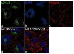
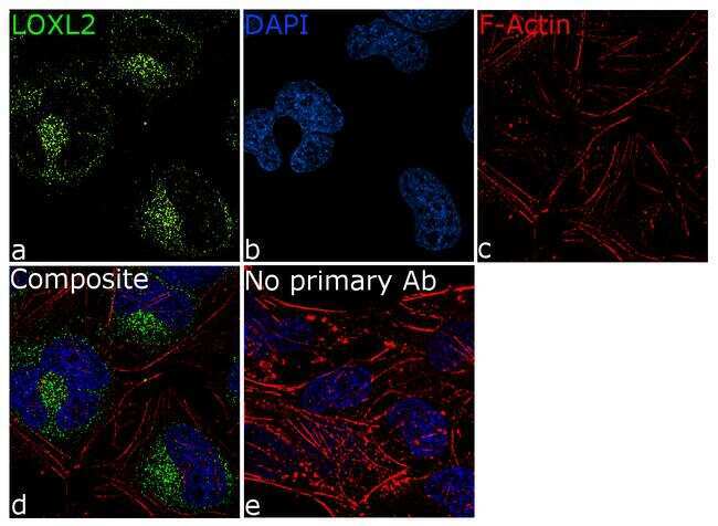
- For immunofluorescence analysis, HeLa cells were fixed and permeabilized for detection of endogenous LOXL2 using Anti-LOXL2 Recombinant Rabbit Monoclonal Antibody (Product # 702694, 5 µg/mL) and labeled with Goat anti-Rabbit IgG (H+L) Superclonal™ Secondary Antibody, Alexa Fluor® 488 conjugate (Product # A27034, 1:2000). Panel a) shows representative cells that were stained for detection and localization of LOXL2 protein (green), Panel b) is stained for nuclei (blue) using SlowFade® Gold Antifade Mountant with DAPI (Product # S36938). Panel c) represents cytoskeletal F-actin staining using Rhodamine Phalloidin (Product # R415, 1:300). Panel d) is a composite image of Panels a, b and c clearly demonstrating perinuclear localization of LOXL2. Panel e) represents control cells with no primary antibody to assess background. The images were captured at 60X magnification.
Supportive validation
- Submitted by
- Invitrogen Antibodies (provider)
- Main image
- Experimental details
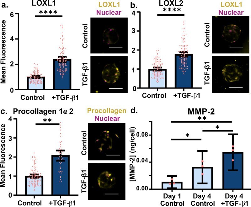
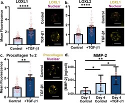
- FIG. 2. Amplified ECM remodeling occurs after fibroblast activation in collagen microtissues. (a) Lysl oxidase like protein 1 and (b) protein 2 (yellow) were stained and quantified. Mean fluorescence values were normalized by cell number [quantified by nuclear count (pink)]. (c) After 4 days of culture, fibroblasts were stained for procollagen 1alpha2 (yellow), a proxy indicating collagen I synthesis, and Hoechst to visualize nuclei and obtain a cell count for normalization. (d) NHLFs encapsulated in microtissues secreted MMP-2, quantified with an ELISA, and normalized by cell number. Experiment performed three times, with the average of each replicate depicted with a circle; SEM (scanning electron microscopy) shown (pink) (scale 100 mu m; points represent individual microtissues from single experiment).