Explore
 Validate
 Learn
Learn Western blot
Antibody data
- Antibody Data
- Antigen structure
- References [0]
- Comments [0]
- Validations
- Western blot [1]
- Immunohistochemistry [5]
- Flow cytometry [1]
Submit
Validation data
Reference
Comment
Report error
- Product number
- 10-7006 - Provider product page

- Provider
- ABEOMICS Inc.
- Product name
- Anti-MBD2/MBD3 Antibody
- Antibody type
- Monoclonal
- Description
- MBD2 is a subunit of the NuRD complex that is postulated to mediate gene repression via recruitment of the complex to methylated DNA. This protein binds around 1 kb downstream of the transcription start site of a subset of 400 CpG island promoters that are characterized by the presence of active histone marks, RNA polymerase II (Pol2) and low to medium gene expression levels and H3K36me3 deposition. Methylated MBD2 is involved in silencing methylated tumor suppressor genes as well as activation of pro-metastatic genes. MBD2 also plays a key role in regulating expression of a range of genes that are associated with optimal DC (Dendritic cells) activation and function.
- Reactivity
- Human
- Host
- Mouse
- Conjugate
- Unconjugated
- Antigen sequence
A partial length recombinant MBD3 p
rotein (amino acids 1-290) was used
 as the immunogen for this antibody
.- Isotype
- IgG
- Antibody clone number
- ABM14A8
- Vial size
- 100 µg
- Concentration
- 0.5 mg/ml
- Storage
- Store the antibody at 4°C, stable for 6 months. For long-term storage, store at -20°C. Avoid repeat freez thawing
No comments: Submit comment
Supportive validation
- Submitted by
- ABEOMICS Inc. (provider)
- Main image

- Experimental details
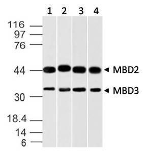
- Western blot analysis of MBD3 (and MBD2). Anti-MBD3 (and MBD2) antibody (Clone: ABM14A8) was used at 2 µg/ml on A431, K562, A375 and HEK293 lysates.
- Protocol
- Protocol
Supportive validation
- Submitted by
- ABEOMICS Inc. (provider)
- Main image
- Experimental details
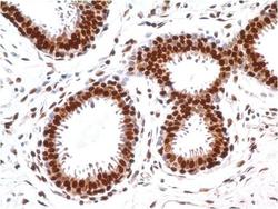
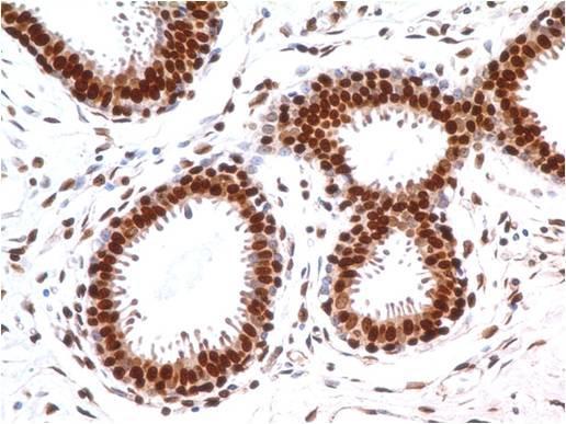
- Immunohistochemical analysis of MBD3 in human breast tissue using MBD3 antibody (Clone: ABM14A8) at 5 µg/ml.
- Protocol
- Protocol
- Submitted by
- ABEOMICS Inc. (provider)
- Main image
- Experimental details
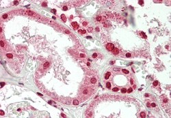
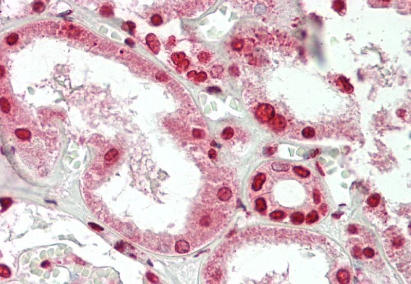
- Immunohistochemical analysis of MBD3 in human Kidney tissue using MBD3 antibody (Clone: ABM14A8) at 10 µg/ml.
- Protocol
- Protocol
- Submitted by
- ABEOMICS Inc. (provider)
- Main image
- Experimental details
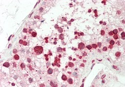
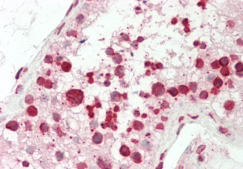
- Immunohistochemical analysis of MBD3 in human Testis tissue using MBD3 antibody (Clone: ABM14A8) at 10 µg/ml.
- Protocol
- Protocol
- Submitted by
- ABEOMICS Inc. (provider)
- Main image
- Experimental details
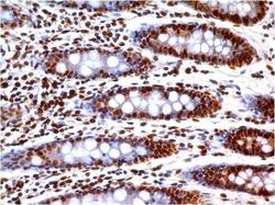
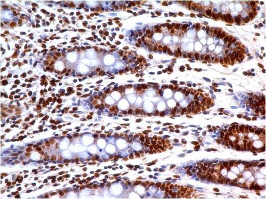
- Immunohistochemical analysis of MBD3 in human Colon tissue using MBD3 antibody (Clone: ABM14A8) at 5 µg/ml.
- Protocol
- Protocol
- Submitted by
- ABEOMICS Inc. (provider)
- Main image
- Experimental details
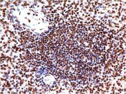
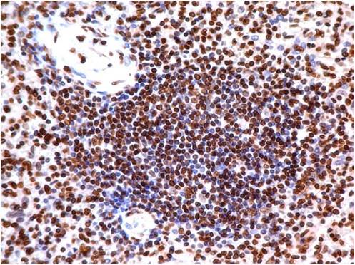
- Immunohistochemical analysis of MBD3 in human Spleen tissue using MBD3 antibody (Clone: ABM14A8) at 5 µg/ml.
- Protocol
- Protocol
Supportive validation
- Submitted by
- ABEOMICS Inc. (provider)
- Main image
- Experimental details
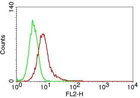
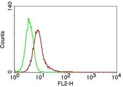
- Intracellular flow analysis of MBD2/MBD3 in Jurkat cells using 0.5 µg/10^6 cells of MBD2/MBD3 antibody (Clone: ABM14A8). Green represents isotype control; red represents anti-MBD2/MBD3 antibody. Goat anti-mouse PE conjugate was used as secondary antibody.
- Protocol
- Protocol