 Explore
 Validate
 Learn
Learn Western blot
 Immunocytochemistry
Antibody data
- Antibody Data
- Antigen structure
- References [2]
- Comments [0]
- Validations
- Immunocytochemistry [5]
- Immunohistochemistry [1]
- Other assay [1]
Submit
Validation data
Reference
Comment
Report error
- Product number
- PA5-29135 - Provider product page

- Provider
- Invitrogen Antibodies
- Product name
- TUBA8 Polyclonal Antibody
- Antibody type
- Polyclonal
- Antigen
- Recombinant full-length protein
- Description
- Recommended positive controls: 293T, A431, H1299, HeLa, HepG2, Molt-4, Raji, NIH-3T3. Predicted reactivity: Mouse (99%), Rat (99%), Xenopus laevis (87%), Chimpanzee (100%), Bovine (98%). Store product as a concentrated solution. Centrifuge briefly prior to opening the vial.
- Reactivity
- Human, Mouse, Rat
- Host
- Rabbit
- Isotype
- IgG
- Vial size
- 100 μL
- Concentration
- 1 mg/mL
- Storage
- Store at 4°C short term. For long term storage, store at -20°C, avoiding freeze/thaw cycles.
Submitted references Human IDO-competent, long-lived immunoregulatory dendritic cells induced by intracellular pathogen, and their fate in humanized mice.
Nanostructured lipid carrier mediates effective delivery of methotrexate to induce apoptosis of rheumatoid arthritis via NF-κB and FOXO1.
Tyagi RK, Miles B, Parmar R, Garg NK, Dalai SK, Baban B, Cutler CW
Scientific reports 2017 Feb 15;7:41083
Scientific reports 2017 Feb 15;7:41083
Nanostructured lipid carrier mediates effective delivery of methotrexate to induce apoptosis of rheumatoid arthritis via NF-κB and FOXO1.
Garg NK, Tyagi RK, Singh B, Sharma G, Nirbhavane P, Kushwah V, Jain S, Katare OP
International journal of pharmaceutics 2016 Feb 29;499(1-2):301-320
International journal of pharmaceutics 2016 Feb 29;499(1-2):301-320
No comments: Submit comment
Supportive validation
- Submitted by
- Invitrogen Antibodies (provider)
- Main image
- Experimental details
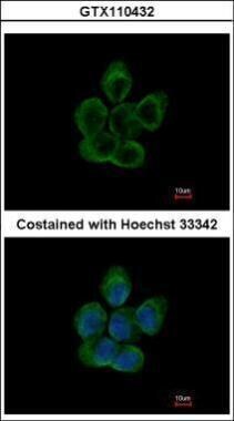
- Immunofluorescent analysis of alpha Tubulin 8 in paraformaldehyde-fixed A431 cells using an Alpha Tubulin 8 polyclonal antibody (Product # PA5-29135) at a 1:200 dilution.

- Submitted by
- Invitrogen Antibodies (provider)
- Main image
- Experimental details
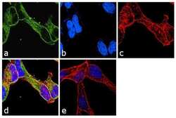
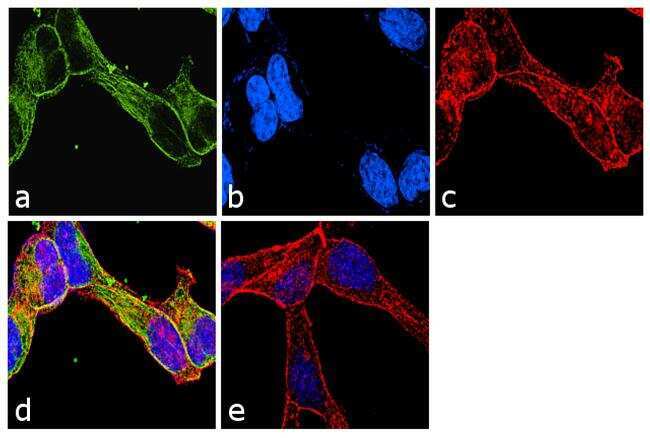
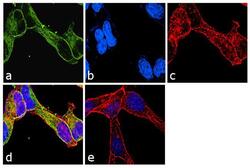
- Immunofluorescence analysis of TUBA8 was performed using 70% confluent log phase LNCaP cells. The cells were fixed with 4% paraformaldehyde for 10 minutes, permeabilized with 0.1% Triton™ X-100 for 10 minutes, and blocked with 1% BSA for 1 hour at room temperature. The cells were labeled with TUBA8 Rabbit Polyclonal Antibody (Product # PA5-29135) at 2 µg/mL in 0.1% BSA and incubated for 3 hours at room temperature and then labeled with Goat anti-Rabbit IgG (H+L) Superclonal™ Secondary Antibody, Alexa Fluor® 488 conjugate (Product # A27034) at a dilution of 1:2000 for 45 minutes at room temperature (Panel a: green). Nuclei (Panel b: blue) were stained with SlowFade® Gold Antifade Mountant with DAPI (Product # S36938). F-actin (Panel c: red) was stained with Rhodamine Phalloidin (Product # R415, 1:300). Panel d represents the merged image showing cytoplasmic localization. Panel e shows the no primary antibody control. The images were captured at 60X magnification.
- Submitted by
- Invitrogen Antibodies (provider)
- Main image

- Experimental details
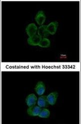
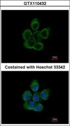
- Immunocytochemistry-Immunofluorescence analysis of TUBA8 in paraformaldehyde-fixed A431 cells using TUBA8 Polyclonal Antibody (Product # PA5-29135) at a dilution of 1:200.
- Submitted by
- Invitrogen Antibodies (provider)
- Main image
- Experimental details
- Immunocytochemistry-Immunofluorescence analysis of TUBA8 in paraformaldehyde-fixed A431 cells using TUBA8 Polyclonal Antibody (Product # PA5-29135) at a dilution of 1:200.

- Submitted by
- Invitrogen Antibodies (provider)
- Main image
- Experimental details
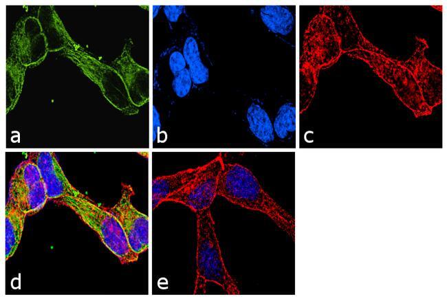
- Immunofluorescence analysis of TUBA8 was performed using 70% confluent log phase LNCaP cells. The cells were fixed with 4% paraformaldehyde for 10 minutes, permeabilized with 0.1% Triton™ X-100 for 10 minutes, and blocked with 1% BSA for 1 hour at room temperature. The cells were labeled with TUBA8 Rabbit Polyclonal Antibody (Product # PA5-29135) at 2 µg/mL in 0.1% BSA and incubated for 3 hours at room temperature and then labeled with Goat anti-Rabbit IgG (Heavy Chain) Superclonal™ Secondary Antibody, Alexa Fluor® 488 conjugate (Product # A27034) at a dilution of 1:2000 for 45 minutes at room temperature (Panel a: green). Nuclei (Panel b: blue) were stained with SlowFade® Gold Antifade Mountant with DAPI (Product # S36938). F-actin (Panel c: red) was stained with Rhodamine Phalloidin (Product # R415, 1:300). Panel d represents the merged image showing cytoplasmic localization. Panel e shows the no primary antibody control. The images were captured at 60X magnification.
Supportive validation
- Submitted by
- Invitrogen Antibodies (provider)
- Main image

- Experimental details
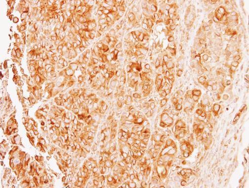
- Immunohistochemical analysis of paraffin-embedded SW480 xenograft, using alpha Tubulin 8 (Product # PA5-29135) antibody at 1:500 dilution. Antigen Retrieval: Citrate buffer, pH 6.0, 15 min.
Supportive validation
- Submitted by
- Invitrogen Antibodies (provider)
- Main image
- Experimental details
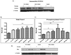
- Figure 2 Phosphorylated Foxo1 decreases with PDDC apoptosis rate. ( A ) FOXO1 expression was observed on protein level in PDDCs (WT & DPG) by carrying out Western blot using Anti-FOXO1 Mab. alpha-Tubulin, loading control (n = 2). The amount of total PDDC Foxo1 ( B ) and phosphorylated Foxo1 ( C ) were measured by whole cell ELISA and normalized to total cell numbers (n = 3) (B) PDDCs generated with E. coli LPS or either of the P. gingivalis strains have significantly higher levels of Foxo1. ( C ) PDDCs generated by the minor-fimbriae expressing WT and DPG-3 or the non-fimbriated MFB have significantly higher levels of phosphorylated Foxo1 ( D ) DCs were washed in RPMI and then incubated for 10 hrs in 10% FBS in RPMI. DCs were then lysed and Bim was detected by immunoblot. alpha-tubulin, loading control (n = 2).
