Explore
 Validate
 Learn
Learn Western blot
 Immunohistochemistry
Antibody data
- Antibody Data
- Antigen structure
- References [1]
- Comments [0]
- Validations
- Immunohistochemistry [1]
- Other assay [1]
Submit
Validation data
Reference
Comment
Report error
- Product number
- PA5-77793 - Provider product page

- Provider
- Invitrogen Antibodies
- Product name
- HSP105 Polyclonal Antibody
- Antibody type
- Polyclonal
- Antigen
- Synthetic peptide
- Description
- Immunohistochemistry samples may be prepared using Bouin's Fixative solution. 1 µg/mL of PA5-77793 was sufficient for detection of HSP110 in 10 µg of human cell line mixed lysate by colorimetric immunoblot analysis using Goat anti-rabbit IgG:HRP as the secondary antibody.|Detects approximately 110kDa.
- Reactivity
- Human, Mouse, Rat, Bovine, Hamster, Yeast
- Host
- Rabbit
- Isotype
- IgG
- Vial size
- 100 µg
- Concentration
- 1 mg/mL
- Storage
- -20°C
Submitted references Embryonic Trophectoderm Secretomics Reveals Chemotactic Migration and Intercellular Communication of Endometrial and Circulating MSCs in Embryonic Implantation.
Calle A, Toribio V, Yáñez-Mó M, Ramírez MÁ
International journal of molecular sciences 2021 May 26;22(11)
International journal of molecular sciences 2021 May 26;22(11)
No comments: Submit comment
Supportive validation
- Submitted by
- Invitrogen Antibodies (provider)
- Main image
- Experimental details
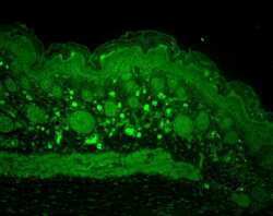
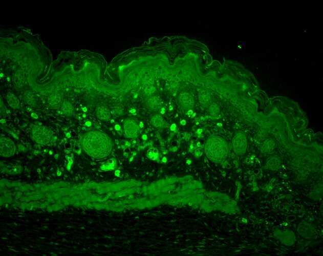
- Immunohistochemistry analysis of HSPH1 in mouse back skin. Samples were incubated with HSPH1 polyclonal antibody (Product # PA5-77793) using a dilution of 1:100 (1 hour at RT) and followed by FITC Goat Anti-Rabbit (green) at a dilution of 1:50 (1 hour at RT).
Supportive validation
- Submitted by
- Invitrogen Antibodies (provider)
- Main image

- Experimental details
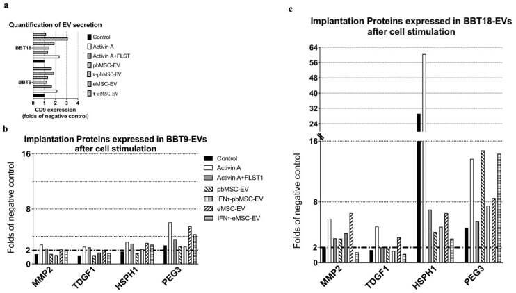
- Figure 6 BBT secret implantation proteins on EVs, and their secretion pattern, which is altered after the uptake of MSC-EVs or the presence of uterine cytokines. SEC-elution profile of EVs from BBT-9 and BBT-18 for the different experimental conditions (Control, Activin A, Activin A+FLST, pbMSC-EVs, tau-pbMSC-EVs, eMSC-EVs, and tau-eMSC-EVs) were analyzed by Dot Blot using an anti-CD9 specific antibody. The positive EV fractions of each experimental condition were quantified by ImageJ software analysis of the dot blot results ( a ). Bead-assisted flow cytometry analysis of selected implantation proteins expressed in BBT-9- ( b ) and BBT-18-EVs ( c ) after cell stimulation.