 Explore
 Validate
 Learn
Learn Western blot
 ELISA
ELISAAntibody data
- Antibody Data
- Antigen structure
- References [6]
- Comments [0]
- Validations
- ELISA [2]
- Immunocytochemistry [1]
Submit
Validation data
Reference
Comment
Report error
- Product number
- BAF1729 - Provider product page

- Provider
- R&D Systems
- Product name
- Mouse TREM2 Biotinylated Antibody
- Antibody type
- Polyclonal
- Description
- Antigen Affinity-purified. Detects mouse TREM2 in Western blots. In Western blots, approximately 5% cross-reactivity with recombinant human (rh) TREM2 is observed and less than 1% cross-reactivity with recombinant mouse (rm) TREM1 and rmTREM3 is observed.
- Reactivity
- Mouse
- Host
- Sheep
- Conjugate
- Biotin
- Antigen sequence
Q99NH8- Isotype
- IgG
- Vial size
- 50 ug
- Concentration
- LYOPH
- Storage
- Use a manual defrost freezer and avoid repeated freeze-thaw cycles. 12 months from date of receipt, -20 to -70 °C as supplied. 1 month, 2 to 8 °C under sterile conditions after reconstitution. 6 months, -20 to -70 °C under sterile conditions after reconstitution.
Submitted references Soluble TREM2 ameliorates pathological phenotypes by modulating microglial functions in an Alzheimer's disease model.
Opposite microglial activation stages upon loss of PGRN or TREM2 result in reduced cerebral glucose metabolism.
Increase of TREM2 during Aging of an Alzheimer's Disease Mouse Model Is Paralleled by Microglial Activation and Amyloidosis.
The FTD-like syndrome causing TREM2 T66M mutation impairs microglia function, brain perfusion, and glucose metabolism.
The triggering receptor expressed on myeloid cells 2 inhibits complement component 1q effector mechanisms and exerts detrimental effects during pneumococcal pneumonia.
DAP12 is required for macrophage recruitment to the lung in response to cigarette smoke and chemotaxis toward CCL2.
Zhong L, Xu Y, Zhuo R, Wang T, Wang K, Huang R, Wang D, Gao Y, Zhu Y, Sheng X, Chen K, Wang N, Zhu L, Can D, Marten Y, Shinohara M, Liu CC, Du D, Sun H, Wen L, Xu H, Bu G, Chen XF
Nature communications 2019 Mar 25;10(1):1365
Nature communications 2019 Mar 25;10(1):1365
Opposite microglial activation stages upon loss of PGRN or TREM2 result in reduced cerebral glucose metabolism.
Götzl JK, Brendel M, Werner G, Parhizkar S, Sebastian Monasor L, Kleinberger G, Colombo AV, Deussing M, Wagner M, Winkelmann J, Diehl-Schmid J, Levin J, Fellerer K, Reifschneider A, Bultmann S, Bartenstein P, Rominger A, Tahirovic S, Smith ST, Madore C, Butovsky O, Capell A, Haass C
EMBO molecular medicine 2019 Jun;11(6)
EMBO molecular medicine 2019 Jun;11(6)
Increase of TREM2 during Aging of an Alzheimer's Disease Mouse Model Is Paralleled by Microglial Activation and Amyloidosis.
Brendel M, Kleinberger G, Probst F, Jaworska A, Overhoff F, Blume T, Albert NL, Carlsen J, Lindner S, Gildehaus FJ, Ozmen L, Suárez-Calvet M, Bartenstein P, Baumann K, Ewers M, Herms J, Haass C, Rominger A
Frontiers in aging neuroscience 2017;9:8
Frontiers in aging neuroscience 2017;9:8
The FTD-like syndrome causing TREM2 T66M mutation impairs microglia function, brain perfusion, and glucose metabolism.
Kleinberger G, Brendel M, Mracsko E, Wefers B, Groeneweg L, Xiang X, Focke C, Deußing M, Suárez-Calvet M, Mazaheri F, Parhizkar S, Pettkus N, Wurst W, Feederle R, Bartenstein P, Mueggler T, Arzberger T, Knuesel I, Rominger A, Haass C
The EMBO journal 2017 Jul 3;36(13):1837-1853
The EMBO journal 2017 Jul 3;36(13):1837-1853
The triggering receptor expressed on myeloid cells 2 inhibits complement component 1q effector mechanisms and exerts detrimental effects during pneumococcal pneumonia.
Sharif O, Gawish R, Warszawska JM, Martins R, Lakovits K, Hladik A, Doninger B, Brunner J, Korosec A, Schwarzenbacher RE, Berg T, Kralovics R, Colinge J, Mesteri I, Gilfillan S, Salmaggi A, Verschoor A, Colonna M, Knapp S
PLoS pathogens 2014 Jun;10(6):e1004167
PLoS pathogens 2014 Jun;10(6):e1004167
DAP12 is required for macrophage recruitment to the lung in response to cigarette smoke and chemotaxis toward CCL2.
Koth LL, Cambier CJ, Ellwanger A, Solon M, Hou L, Lanier LL, Abram CL, Hamerman JA, Woodruff PG
Journal of immunology (Baltimore, Md. : 1950) 2010 Jun 1;184(11):6522-8
Journal of immunology (Baltimore, Md. : 1950) 2010 Jun 1;184(11):6522-8
No comments: Submit comment
Supportive validation
- Submitted by
- R&D Systems (provider)
- Main image

- Experimental details
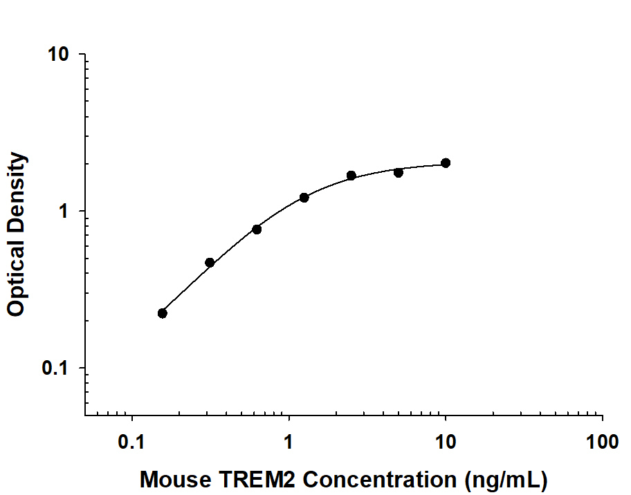
- Mouse TREM2 ELISA Standard Curve. Recombinant Human/Mouse TREM2 protein was serially diluted 2-fold and captured by Rat Anti-Human/Mouse TREM2 Monoclonal Antibody (Catalog # MAB17291) coated on a Clear Polystyrene Microplate (Catalog # DY990). Sheep Anti-Mouse TREM2 Biotinylated Antigen Affinity-purified Polyclonal Antibody (Catalog # BAF1729) was incubated with the protein captured on the plate. Detection of the standard curve was achieved by incubating Streptavidin-HRP (Catalog # DY998) followed by Substrate Solution (Catalog # DY999) and stopping the enzymatic reaction with Stop Solution (Catalog # DY994).
- Submitted by
- R&D Systems (provider)
- Main image
- Experimental details
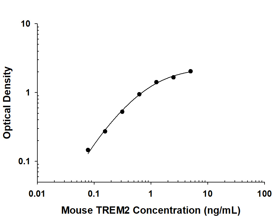
- Mouse TREM2 ELISA Standard Curve. Recombinant Mouse TREM2 protein was serially diluted 2-fold and captured by Sheep Anti-Mouse TREM2 Antigen Affinity-purified Polyclonal Antibody (Catalog # AF1729) coated on a Clear Polystyrene Microplate (Catalog # DY990). Sheep Anti-Mouse TREM2 Biotinylated Antigen Affinity-purified Polyclonal Antibody (Catalog # BAF1729) was incubated with the protein captured on the plate. Detection of the standard curve was achieved by incubating Streptavidin-HRP (Catalog # DY998) followed by Substrate Solution (Catalog # DY999) and stopping the enzymatic reaction with Stop Solution (Catalog # DY994).
Supportive validation
- Submitted by
- R&D Systems (provider)
- Main image
- Experimental details
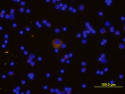
- TREM-2 in Mouse Splenocytes. TREM-2 was detected in immersion fixed mouse spleno-cytes using Mouse Anti-TREM-2 Biotinylated Antigen Affinity-purified Polyclonal Antibody (Catalog # BAF1729) at 10 µg/mL for 3 hours at room temperature. Cells were stained using the NorthernLights™ 557-conjugated Streptavidin (yellow; Catalog # NL999) and counterstained with DAPI (blue). View our protocol for Fluorescent ICC Staining of Non-adherent Cells.
