Explore
 Validate
 Learn
Learn Western blot
 Flow cytometry
Antibody data
- Antibody Data
- Antigen structure
- References [0]
- Comments [0]
- Validations
- Western blot [2]
- Immunohistochemistry [1]
Submit
Validation data
Reference
Comment
Report error
- Product number
- PA5-46980 - Provider product page

- Provider
- Invitrogen Antibodies
- Product name
- Anti-TREM2 Polyclonal Antibody
- Antibody type
- Polyclonal
- Antigen
- Recombinant full-length protein
- Description
- In direct ELISAs, approximately 5% cross-reactivity with recombinant mouse (rm) TREM-2b is observed and less than 1% cross-reactivity with recombinant human (rh) TREM-1, rhTLT-1, rmTREM-L2, rmTREM-3 and rmTREM-4 is observed. Reconstitute at 0.2 mg/mL in sterile PBS.
- Reactivity
- Human
- Host
- Goat
- Isotype
- IgG
- Vial size
- 100 µg
- Concentration
- 0.2 mg/mL
- Storage
- -20° C, Avoid Freeze/Thaw Cycles
No comments: Submit comment
Supportive validation
- Submitted by
- Invitrogen Antibodies (provider)
- Main image
- Experimental details
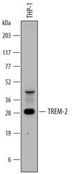
- Western blot analysis from lysates of THP-1 human acute monocytic leukemia cell line. PVDF membrane was probed with 0.2 µg/mL of Goat Anti-human TREM-2 Antigen Affinity-purified Polyclonal Antibody (Product # PA5-46980) followed by HRP-conjugated Anti-Goat IgG Secondary Antibody. A specific band was detected for TREM-2 at approximately 28 kDa (as indicated). This experiment was conducted under reducing conditions.

- Submitted by
- Invitrogen Antibodies (provider)
- Main image
- Experimental details
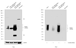
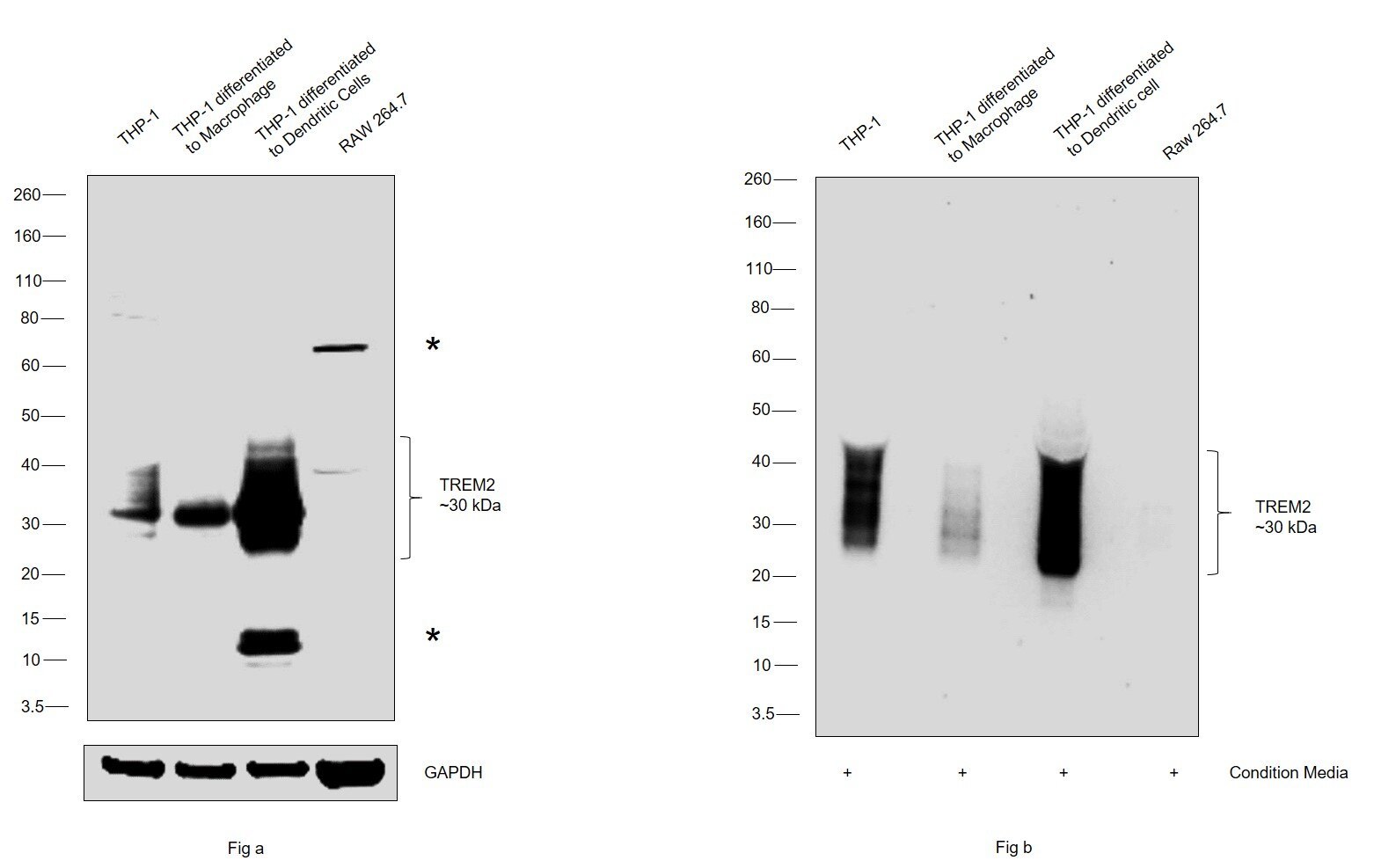
- Western blot was performed using TREM2 Polyclonal Antibody (Product # PA5-46980) and a 30 kDa band corresponding to TREM2 was observed in cell lines THP-1, THP-1 differentiated to Macrophage, THP-1 differentiated to Dendritic cells and condition media of THP-1, THP-1 differentiated to Macrophage, THP-1 differentiated to Dendritic cells; and was absent in RAW264.7 and condition media of Raw 264.7 along with two uncharacterized bands (*) 60 kDa and 12 kDa. Whole cell extracts (30 µg lysate) of THP-1 (Lane 1), THP-1 differentiated to Macrophage (Lane 2), THP-1 differentiated to Dendritic cells (Lane 3), RAW264.7 (Lane 4) of Fig a; and condition media of THP-1 (Lane 1), THP-1 differentiated to Macrophage (Lane 2), THP-1 differentiated to Dendritic cells (Lane 3), RAW264.7 (Lane 4) of Fig b were electrophoresed using NuPAGE™ 4-12% Bis-Tris Protein Gel (Product # NP0321BOX). Resolved proteins were then transferred onto a Nitrocellulose membrane (Product # LC2001) by iBlot® 2 Dry Blotting System (Product # IB21001). The blot was probed with the primary antibody (0.2 ug/ml) and detected by chemiluminescence with Rabbit anti-Goat IgG (H+L) Superclonal™ Recombinant Secondary Antibody, HRP (Product # A27014, 1:4000 dilution) using the iBright FL 1000 (Product # A32752). Chemiluminescent detection was performed using Novex® ECL Chemiluminescent Substrate Reagent Kit (Product # WP20005).
Supportive validation
- Submitted by
- Invitrogen Antibodies (provider)
- Main image
- Experimental details
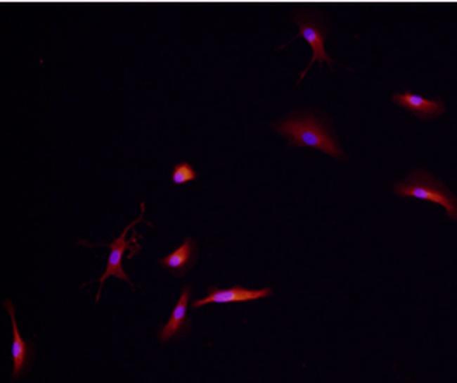
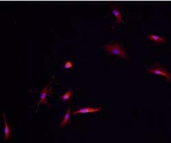
- Immunocytochemical analysis of TREM-2 was detected in immersion fixed immature human dendritic cells using Goat Anti-human TREM-2 Antigen Affinity-purified Polyclonal Antibody (Product # PA5-46980) at 10 µg/mL for 3 hours at room temperature. Cells were stained using the 557-conjugated Anti-Goat IgG Secondary Antibody (re and counterstained with DAPI (blue). Specific staining was localized to cytoplasm.