Explore
 Validate
 Learn
LearnMA5-29587
antibody from Invitrogen Antibodies
Targeting: STUB1
CHIP, HSPABP2, NY-CO-7, SDCCAG7, UBOX1
 Western blot
 ELISA
ELISAAntibody data
- Antibody Data
- Antigen structure
- References [0]
- Comments [0]
- Validations
- Western blot [2]
- Immunocytochemistry [4]
- Immunoprecipitation [1]
- Flow cytometry [2]
- Other assay [1]
Submit
Validation data
Reference
Comment
Report error
- Product number
- MA5-29587 - Provider product page

- Provider
- Invitrogen Antibodies
- Product name
- STUB1 Recombinant Rabbit Monoclonal Antibody (034)
- Antibody type
- Monoclonal
- Antigen
- Recombinant full-length protein
- Description
- This product is preservative free. It is recommended to add sodium azide to avoid contamination (final concentration 0.05%-0.1%). Recombinant rabbit monoclonal antibodies are produced using in vitro expression systems. The expression systems are developed by cloning in the specific antibody DNA sequences from immunoreactive rabbits. Then, individual clones are screened to select the best candidates for production. The advantages of using recombinant rabbit monoclonal antibodies include: better specificity and sensitivity, lot-to-lot consistency, animal origin-free formulations, and broader immunoreactivity to diverse targets due to larger rabbit immune repertoire. This antibody has specificity for Human STUB1.
- Reactivity
- Human
- Host
- Rabbit
- Isotype
- IgG
- Antibody clone number
- 34
- Vial size
- 100 μL
- Concentration
- 1 mg/mL
- Storage
- Store at 4°C short term. For long term storage, store at -20°C, avoiding freeze/thaw cycles.
No comments: Submit comment
Supportive validation
- Submitted by
- Invitrogen Antibodies (provider)
- Main image
- Experimental details
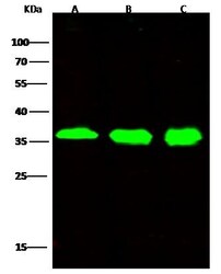
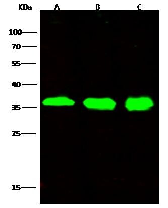
- Western Blot using STUB1 Recombinant Rabbit Monoclonal Antibody (34) (Product # MA5-29587) at 1:500 dilution. Lane A: HELA Whole Cell Lysate, Lane B: 293T Whole Cell Lysate, Lane C: MCF7 Whole Cell Lysate. Lysates/proteins at 30 μg per lane. Secondary Goat Anti-Rabbit IgG H&L (DyLight™ 800) at 1:10,000 dilution. Developed using the Odyssey technique. Performed under reducing conditions. Predicted band size: 35 kDa. Observed band size: 36 kDa.
- Submitted by
- Invitrogen Antibodies (provider)
- Main image
- Experimental details
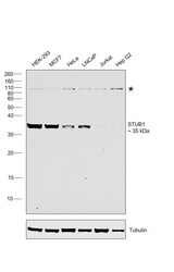
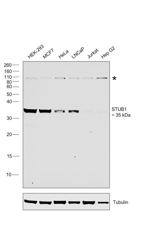
- Western blot was performed using Anti-E3 ubiquitin-protein ligase CHIP (STUB1) Monoclonal Antibody (Product # MA5-29587) on whole cell extracts (30 µg lysate) of HEK-293 (Lane 1), MCF7 (Lane 2), HeLa (Lane 3), LNCaP (Lane 4), Jurkat (Lane 5) and HepG2 (Lane 6) and 35 kDa band corresponding to STUB1 was observed along with an uncharacterized band (*) at ~110 kDa. Resolved proteins were then transferred onto a nitrocellulose membrane (Product # IB23001) by iBlot® 2 Dry Blotting System (Product # IB21001). The blot was probed with the primary antibody (1:1,000 dilution) and detected by Goat anti-Rabbit IgG (Heavy Chain) Superclonal™ Recombinant Secondary Antibody, HRP conjugate (Product # A27036, 0.25 µg/mL, 1:4,000 dilution) using the iBright FL 1000 (Product # A32752). Chemiluminescent detection was performed using Novex® ECL Chemiluminescent Substrate Reagent Kit (Product # WP20005).
Supportive validation
- Submitted by
- Invitrogen Antibodies (provider)
- Main image
- Experimental details
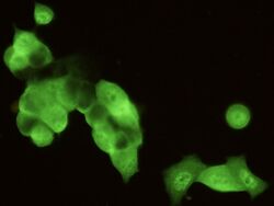
- Immunofluorescence staining of STUB1 in MCF7 cells. Cells were fixed with 4% PFA, permeabilzed with 0.1% Triton X-100 in PBS, blocked with 10% serum, and incubated with STUB1 Recombinant Rabbit Monoclonal Antibody (34) (Product # MA5-29587, 1:300) at 4°C overnight. Then cells were stained with the Alexa Fluor®488-conjugated Goat Anti-rabbit IgG secondary antibody (green). Positive staining was localized to cytoplasm and nucleus.

- Submitted by
- Invitrogen Antibodies (provider)
- Main image

- Experimental details
- Immunofluorescence analysis of STUB1 was performed using 70% confluent log phase MCF7 cells. The cells were fixed with 4% paraformaldehyde for 10 minutes, permeabilized with 0.1% Triton™ X-100 for 15 minutes, and blocked with 2% BSA for 1 hour at room temperature. The cells were labeled with STUB1 Recombinant Rabbit Monoclonal Antibody (Product # MA5-29587) at 1:100 dilution in 0.1% BSA, incubated at 4 degree Celsius overnight and then labeled with Goat anti-Rabbit IgG (H+L) Superclonal™ Recombinant Secondary Antibody, Alexa Fluor® 488 conjugate (Product # A27034) at a dilution of 1:2000 for 45 minutes at room temperature (Panel a: green). Nuclei (Panel b: blue) were stained with ProLong™ Diamond Antifade Mountant with DAPI (Product # P36962). F-actin (Panel c: red) was stained with Rhodamine Phalloidin (Product # R415). Panel d represents the merged image showing Nucleus and Cytoplasm localization. Panel e represents control cells with no primary antibody to assess background. The images were captured at 60X magnification.

- Submitted by
- Invitrogen Antibodies (provider)
- Main image

- Experimental details
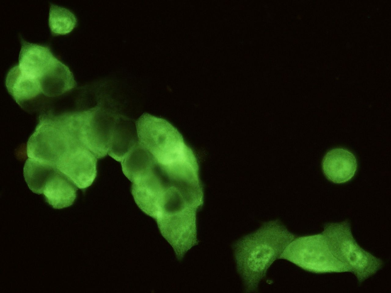
- Immunofluorescence staining of STUB1 in MCF7 cells. Cells were fixed with 4% PFA, permeabilzed with 0.1% Triton X-100 in PBS, blocked with 10% serum, and incubated with STUB1 Recombinant Rabbit Monoclonal Antibody (34) (Product # MA5-29587, 1:300) at 4°C overnight. Then cells were stained with the Alexa Fluor®488-conjugated Goat Anti-rabbit IgG secondary antibody (green). Positive staining was localized to cytoplasm and nucleus.
- Submitted by
- Invitrogen Antibodies (provider)
- Main image

- Experimental details
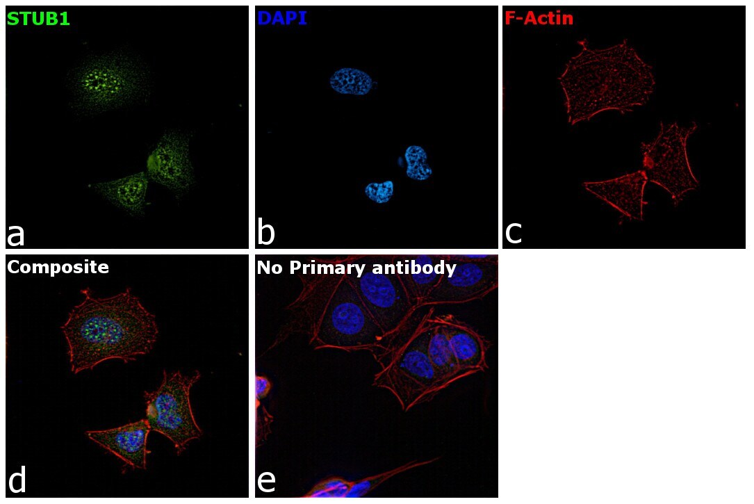
- Immunofluorescence analysis of STUB1 was performed using 70% confluent log phase MCF7 cells. The cells were fixed with 4% paraformaldehyde for 10 minutes, permeabilized with 0.1% Triton™ X-100 for 15 minutes, and blocked with 2% BSA for 1 hour at room temperature. The cells were labeled with STUB1 Recombinant Rabbit Monoclonal Antibody (Product # MA5-29587) at 1:100 dilution in 0.1% BSA, incubated at 4 degree Celsius overnight and then labeled with Goat anti-Rabbit IgG (Heavy Chain) Superclonal™ Recombinant Secondary Antibody, Alexa Fluor® 488 conjugate (Product # A27034) at a dilution of 1:2000 for 45 minutes at room temperature (Panel a: green). Nuclei (Panel b: blue) were stained with ProLong™ Diamond Antifade Mountant with DAPI (Product # P36962). F-actin (Panel c: red) was stained with Rhodamine Phalloidin (Product # R415). Panel d represents the merged image showing Nucleus and Cytoplasm localization. Panel e represents control cells with no primary antibody to assess background. The images were captured at 60X magnification.
Supportive validation
- Submitted by
- Invitrogen Antibodies (provider)
- Main image

- Experimental details
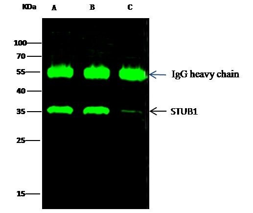
- STUB1 Immunoprecipitation using: Lane A: 0.5 mg Hela Whole Cell Lysate, Lane B: 0.5 mg 293T Whole Cell Lysate, Lane C: 0.5 mg MCF-7 Whole Cell Lysate 2 µL with STUB1 Recombinant Rabbit Monoclonal Antibody (34) (Product # MA5-29587) and 15 µL of 50 % Protein G agarose. Primary antibody: STUB1 Recombinant Rabbit Monoclonal Antibody (34), at 1:500 dilution. Secondary antibody: Dylight 800-labeled antibody to rabbit IgG (H+L), at 1:5,000 dilution. Developed using the Odyssey technique. Performed under reducing conditions. Predicted band size: 35 kDa. Observed band size: 35 kDa.

Supportive validation
- Submitted by
- Invitrogen Antibodies (provider)
- Main image

- Experimental details
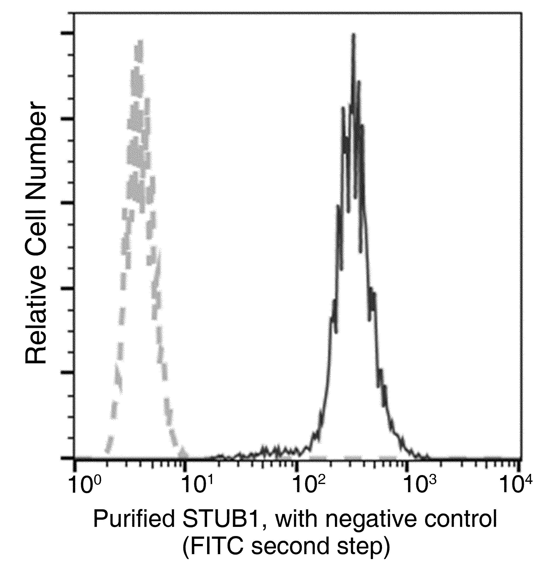
- Flow cytometric analysis of Human STUB1 expression on Hela cells. The cells were treated according to manufacturer’s manual, stained with STUB1 Recombinant Rabbit Monoclonal Antibody (34) (Product # MA5-29587), then a FITC-conjugated Secondary antibody. The fluorescence histograms were derived from gated events with the forward and side light-scatter characteristics of intact cells.

- Submitted by
- Invitrogen Antibodies (provider)
- Main image

- Experimental details
- Flow cytometric analysis of Human STUB1 expression on Hela cells. The cells were treated according to manufacturer’s manual, stained with STUB1 Recombinant Rabbit Monoclonal Antibody (34) (Product # MA5-29587), then a FITC-conjugated Secondary antibody. The fluorescence histograms were derived from gated events with the forward and side light-scatter characteristics of intact cells.
Supportive validation
- Submitted by
- Invitrogen Antibodies (provider)
- Main image

- Experimental details
- STUB1 Immunoprecipitation using: Lane A: 0.5 mg Hela Whole Cell Lysate, Lane B: 0.5 mg 293T Whole Cell Lysate, Lane C: 0.5 mg MCF-7 Whole Cell Lysate 2 µL with STUB1 Recombinant Rabbit Monoclonal Antibody (34) (Product # MA5-29587) and 15 µL of 50 % Protein G agarose. Primary antibody: STUB1 Recombinant Rabbit Monoclonal Antibody (34), at 1:500 dilution. Secondary antibody: Dylight 800-labeled antibody to rabbit IgG (H+L), at 1:5,000 dilution. Developed using the Odyssey technique. Performed under reducing conditions. Predicted band size: 35 kDa. Observed band size: 35 kDa.