Explore
 Validate
 Learn
Learn Western blot
 Flow cytometry
Antibody data
- Antibody Data
- Antigen structure
- References [0]
- Comments [0]
- Validations
- Flow cytometry [1]
Submit
Validation data
Reference
Comment
Report error
- Product number
- PA1-776 - Provider product page

- Provider
- Invitrogen Antibodies
- Product name
- CSP alpha Polyclonal Antibody
- Antibody type
- Polyclonal
- Antigen
- Synthetic peptide
- Description
- PA1-776 detects Cysteine String Protein Alpha in mouse and rat brain samples. PA1-776 has been successfully used in Western blot procedures. By Western blot, this antibody detects a ~34 kDa protein representing Cysteine String Protein Alpha in rat and mouse brain homogenates. The PA1-776 immunogen is a synthetic peptide corresponding to residues E(180) T T Q L T A D S H P S Y H T D G F N(198) of rat CSP. This sequence is conserved across many species including mouse, human and bovine. This peptide (Cat. # PEP-287) is available for use in neutralization and control experiments.
- Reactivity
- Human, Mouse, Rat
- Host
- Rabbit
- Isotype
- IgG
- Vial size
- 100 μg
- Concentration
- 1 mg/mL
- Storage
- -20°C, Avoid Freeze/Thaw Cycles
No comments: Submit comment
Supportive validation
- Submitted by
- Invitrogen Antibodies (provider)
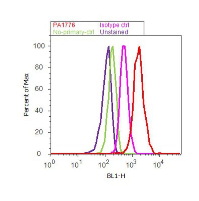
- Main image
- Experimental details
- Flow cytometry analysis of Cysteine String Protein alpha was performed using PC-12 cells. Cells were fixed with 70% ethanol for 10 minutes, permeabilized with 0.25% Triton™ X-100 for 20 minutes, and blocked with 5% BSA for 30 minutes at room temperature. Cells were labeled with Cysteine String Protein alpha Rabbit Polyclonal Antibody (PA1-776, red histogram) or with rabbit isotype control (pink histogram) at 3-5 ug/million cells in 2.5% BSA. After incubation at room temperature for 2 hours, the cells were labeled with Alexa Fluor® 488 Goat Anti-Rabbit Secondary Antibody (A11008) at a dilution of 1:400 for 30 minutes at room temperature. The representative 10,000 cells were acquired and analyzed for each sample using an Attune® Acoustic Focusing Cytometer. The purple histogram represents unstained control cells and the green histogram represents no-primary-antibody control..