Explore
 Validate
 Learn
Learn ELISA
ELISAAntibody data
- Antibody Data
- Antigen structure
- References [0]
- Comments [0]
- Validations
- ELISA [1]
- Immunohistochemistry [1]
Submit
Validation data
Reference
Comment
Report error
- Product number
- MA5-49516 - Provider product page

- Provider
- Invitrogen Antibodies
- Product name
- Cystatin C Monoclonal Antibody (KT145)
- Antibody type
- Monoclonal
- Antigen
- Purifed from natural sources
- Description
- This product was used in both Indirect and Sandwich ELISA.
- Reactivity
- Human
- Host
- Mouse
- Isotype
- IgG
- Antibody clone number
- KT145
- Vial size
- 100 µg
- Concentration
- 1 mg/mL
- Storage
- Store at 4°C short term. For long term storage, store at -20°C, avoiding freeze/thaw cycles.
No comments: Submit comment
Supportive validation
- Submitted by
- Invitrogen Antibodies (provider)
- Main image

- Experimental details
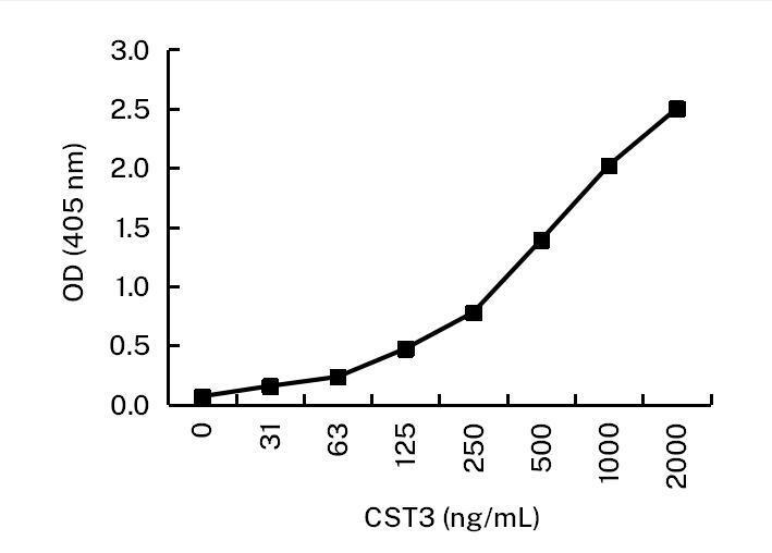
- ELISA of Cystatin C, Microtiter wells were coated with Cystatin C monoclonal antibody (Product # MA5-49516) at 5 µg/mL. Peroxidase conjugated mouse anti-human CST3 monoclonal antibody (Product # MA5-49515) was used as the detection antibody and CST3 was used as the antigen. Result: MA5-49516 and MA5-49515 can be used as a matched antibody pair to detect and quantify the concentration of CST3.
Supportive validation
- Submitted by
- Invitrogen Antibodies (provider)
- Main image
- Experimental details
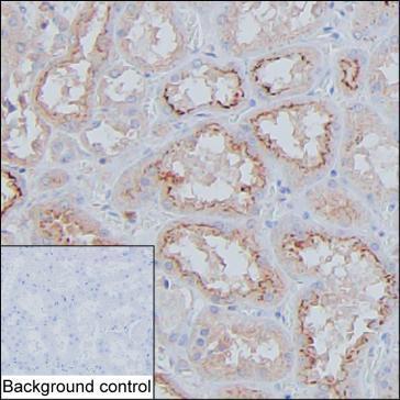
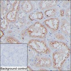
- Immunohistochemistry analysis of Cystatin C in formalin-fixed paraffin-embedded human kidney tissue. Samples were incubated in Cystatin C monoclonal antibody (Product # MA5-49516) using a dilution of 2 µg/mL at room temperature for 1 hour followed by poly-peroxidase conjugated goat anti-mouse IgG secondary antibody. Antigen was retrieved thorough addition of boiling Tris/EDTA buffer pH 9 in a pressure cooker for 3 min. Endogenous peroxidase activity was quenched by incubating the sections with 3% H2O2 for 30 min at room temperature. Diaminobenzidine was used as the chromogen, and the section was counterstained with hematoxylin. A tissue section incubated with phosphate-buffered saline followed by incubation with the secondary antibody was used as the background control. Result: Cells in tubule are positively stained at cytoplasm and cell membrane.