Explore
 Validate
 Learn
Learn Western blot
Antibody data
- Antibody Data
- Antigen structure
- References [2]
- Comments [0]
- Validations
- Western blot [1]
- Immunohistochemistry [1]
Submit
Validation data
Reference
Comment
Report error
- Product number
- MAB5820 - Provider product page

- Provider
- Novus Biologicals
- Product name
- Mouse Monoclonal VAP-A Antibody
- Antibody type
- Monoclonal
- Description
- Protein A or G purified from hybridoma culture supernatant. Detects human VAP-A in direct ELISAs and Western blots. In direct ELISAs, no cross-reactivity with recombinant human VAP-B or recombinant rat VAP-B is observed.
- Reactivity
- Human
- Host
- Mouse
- Isotype
- IgG
- Vial size
- 100 ug
- Concentration
- LYOPH
- Storage
- Use a manual defrost freezer and avoid repeated freeze-thaw cycles. 12 months from date of receipt, -20 to -70 degreesC as supplied. 1 month, 2 to 8 degreesC under sterile conditions after reconstitution. 6 months, -20 to -70 degreesC under sterile conditions after reconstitution.
Submitted references NPC1 regulates ER contacts with endocytic organelles to mediate cholesterol egress.
VAMP associated proteins are required for autophagic and lysosomal degradation by promoting a PtdIns4P-mediated endosomal pathway.
Höglinger D, Burgoyne T, Sanchez-Heras E, Hartwig P, Colaco A, Newton J, Futter CE, Spiegel S, Platt FM, Eden ER
Nature communications 2019 Sep 19;10(1):4276
Nature communications 2019 Sep 19;10(1):4276
VAMP associated proteins are required for autophagic and lysosomal degradation by promoting a PtdIns4P-mediated endosomal pathway.
Mao D, Lin G, Tepe B, Zuo Z, Tan KL, Senturk M, Zhang S, Arenkiel BR, Sardiello M, Bellen HJ
Autophagy 2019 Jul;15(7):1214-1233
Autophagy 2019 Jul;15(7):1214-1233
No comments: Submit comment
Supportive validation
- Submitted by
- Novus Biologicals (provider)
- Main image
- Experimental details
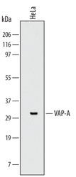
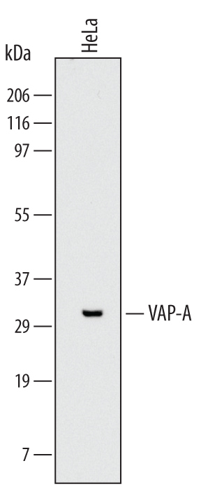
- Detection of Human VAP-A by Western Blot. Western blot shows lysates of HeLa human cervical epithelial carcinoma cell line. PVDF Membrane was probed with 2 µg/mL of Mouse Anti-Human VAP-A Monoclonal Antibody (Catalog # MAB5820) followed by HRP-conjugated Anti-Mouse IgG Secondary Antibody (Catalog # HAF007). A specific band was detected for VAP-A at approximately 33 kDa (as indicated). This experiment was conducted under reducing conditions and using Immunoblot Buffer Group 1 with 0.05% Tween 20.
Supportive validation
- Submitted by
- Novus Biologicals (provider)
- Main image
- Experimental details
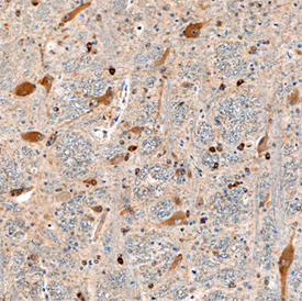
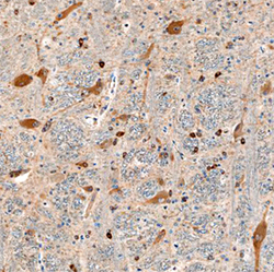
- VAP-A in Human Brain. VAP-A was detected in immersion fixed paraffin-embedded sections of human brain using Mouse Anti-Human VAP-A Monoclonal Antibody (Catalog # MAB5820) at 15 µg/mL overnight at 4 °C. Before incubation with the primary antibody, tissue was subjected to heat-induced epitope retrieval using Antigen Retrieval Reagent-Basic (Catalog # CTS013). Tissue was stained using the Anti-Mouse HRP-DAB Cell & Tissue Staining Kit (brown; Catalog # CTS002) and counterstained with hematoxylin (blue). Specific staining was localized to neuronal cell bodies and processes. View our protocol for Chromogenic IHC Staining of Paraffin-embedded Tissue Sections.