Explore
 Validate
 Learn
Learn Western blot
Antibody data
- Antibody Data
- Antigen structure
- References [4]
- Comments [0]
- Validations
- Western blot [4]
- Immunohistochemistry [2]
Submit
Validation data
Reference
Comment
Report error
- Product number
- NBP1-31237 - Provider product page

- Provider
- Novus Biologicals
- Proper citation
- Novus Cat#NBP1-31237, RRID:AB_2213101
- Product name
- Rabbit Polyclonal VAP-A Antibody
- Antibody type
- Polyclonal
- Description
- Immunogen affinity purified.
- Reactivity
- Human, Mouse, Rat
- Host
- Rabbit
- Isotype
- IgG
- Vial size
- 100 ul
- Storage
- Aliquot and store at -20C or -80C. Avoid freeze-thaw cycles.
Submitted references GRAF2, WDR44, and MICAL1 mediate Rab8/10/11-dependent export of E-cadherin, MMP14, and CFTR ΔF508.
Pitavastatin Differentially Modulates MicroRNA-Associated Cholesterol Transport Proteins in Macrophages.
Extended Synaptotagmin (ESyt) Triple Knock-Out Mice Are Viable and Fertile without Obvious Endoplasmic Reticulum Dysfunction.
Dynamic imaging of the hepatitis C virus NS5A protein during a productive infection.
Lucken-Ardjomande Häsler S, Vallis Y, Pasche M, McMahon HT
The Journal of cell biology 2020 May 4;219(5)
The Journal of cell biology 2020 May 4;219(5)
Pitavastatin Differentially Modulates MicroRNA-Associated Cholesterol Transport Proteins in Macrophages.
Zhang H, Lamon BD, Moran G, Sun T, Gotto AM Jr, Hajjar DP
PloS one 2016;11(7):e0159130
PloS one 2016;11(7):e0159130
Extended Synaptotagmin (ESyt) Triple Knock-Out Mice Are Viable and Fertile without Obvious Endoplasmic Reticulum Dysfunction.
Sclip A, Bacaj T, Giam LR, Südhof TC
PloS one 2016;11(6):e0158295
PloS one 2016;11(6):e0158295
Dynamic imaging of the hepatitis C virus NS5A protein during a productive infection.
Eyre NS, Fiches GN, Aloia AL, Helbig KJ, McCartney EM, McErlean CS, Li K, Aggarwal A, Turville SG, Beard MR
Journal of virology 2014 Apr;88(7):3636-52
Journal of virology 2014 Apr;88(7):3636-52
No comments: Submit comment
Supportive validation
- Submitted by
- Novus Biologicals (provider)
- Main image
- Experimental details
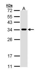
- Western Blot: VAP-A Antibody [NBP1-31237] - Sample (30 ug of whole cell lysate) A:H1299 12% SDS PAGE, antibody diluted at 1:1000.
- Submitted by
- Novus Biologicals (provider)
- Main image

- Experimental details
- Western Blot: VAP-A Antibody [NBP1-31237] - Analysis of antibody on A:NIH-3T3 12% SDS PAGE diluted at 1:1000
- Submitted by
- Novus Biologicals (provider)
- Main image
- Experimental details
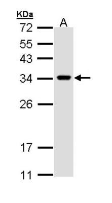
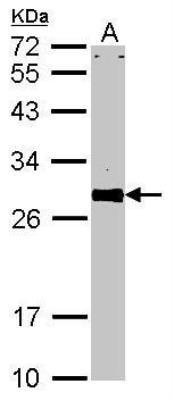
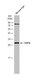
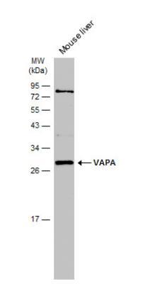
- Western Blot: VAP-A Antibody [NBP1-31237] - Mouse tissue extract (50 ug) was separated by 12% SDS-PAGE, and the membrane was blotted with VAPA antibody [N1N3] diluted at 1:1000. The HRP-conjugated anti-rabbit IgG antibody (NBP2-19301) was used to detect the primary antibody.
- Submitted by
- Novus Biologicals (provider)
- Main image
- Experimental details
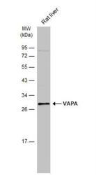
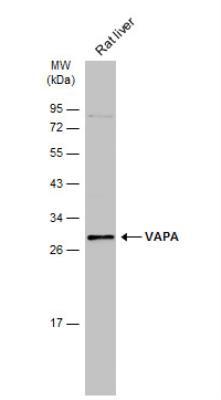
- Western Blot: VAP-A Antibody [NBP1-31237] - Rat tissue extract (50 ug) was separated by 12% SDS-PAGE, and the membrane was blotted with VAPA antibody [N1N3] diluted at 1:1000. The HRP-conjugated anti-rabbit IgG antibody (NBP2-19301) was used to detect the primary antibody.
Supportive validation
- Submitted by
- Novus Biologicals (provider)
- Main image
- Experimental details
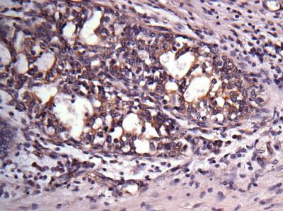
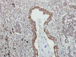
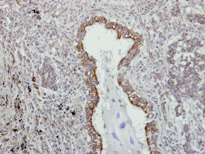
- Immunohistochemistry-Paraffin: VAP-A Antibody [NBP1-31237] - Paraffin-embedded lung AdCA, using antibody at 1:500 dilution.
- Submitted by
- Novus Biologicals (provider)
- Main image
- Experimental details
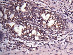
- Immunohistochemistry-Paraffin: VAP-A Antibody [NBP1-31237] - Paraffin-embedded breast ca, using antibody at 1:500 dilution.