Explore
 Validate
 Learn
Learn Western blot
Antibody data
- Antibody Data
- Antigen structure
- References [1]
- Comments [0]
- Validations
- Western blot [1]
- Immunohistochemistry [1]
Submit
Validation data
Reference
Comment
Report error
- Product number
- MAB5820 - Provider product page

- Provider
- R&D Systems
- Product name
- Human VAP-A Antibody
- Antibody type
- Monoclonal
- Description
- Protein A or G purified from hybridoma culture supernatant. Detects human VAP-A in direct ELISAs and Western blots. In direct ELISAs, no cross-reactivity with recombinant human VAP-B or recombinant rat VAP-B is observed.
- Reactivity
- Human
- Host
- Mouse
- Conjugate
- Unconjugated
- Antigen sequence
Q9P0L0- Isotype
- IgG
- Antibody clone number
- 604101
- Vial size
- 100 ug
- Concentration
- LYOPH
- Storage
- Use a manual defrost freezer and avoid repeated freeze-thaw cycles. 12 months from date of receipt, -20 to -70 °C as supplied. 1 month, 2 to 8 °C under sterile conditions after reconstitution. 6 months, -20 to -70 °C under sterile conditions after reconstitution.
Submitted references VAMP associated proteins are required for autophagic and lysosomal degradation by promoting a PtdIns4P-mediated endosomal pathway.
Mao D, Lin G, Tepe B, Zuo Z, Tan KL, Senturk M, Zhang S, Arenkiel BR, Sardiello M, Bellen HJ
Autophagy 2019 Jul;15(7):1214-1233
Autophagy 2019 Jul;15(7):1214-1233
No comments: Submit comment
Supportive validation
- Submitted by
- R&D Systems (provider)
- Main image
- Experimental details
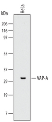
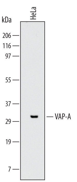
- Detection of Human VAP-A by Western Blot. Western blot shows lysates of HeLa human cervical epithelial carcinoma cell line. PVDF Membrane was probed with 2 µg/mL of Mouse Anti-Human VAP-A Monoclonal Antibody (Catalog # MAB5820) followed by HRP-conjugated Anti-Mouse IgG Secondary Antibody (Catalog # HAF007). A specific band was detected for VAP-A at approximately 33 kDa (as indicated). This experiment was conducted under reducing conditions and using Immunoblot Buffer Group 1 with 0.05% Tween 20.
Supportive validation
- Submitted by
- R&D Systems (provider)
- Main image
- Experimental details
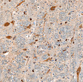
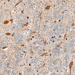
- VAP-A in Human Brain. VAP-A was detected in immersion fixed paraffin-embedded sections of human brain using Mouse Anti-Human VAP-A Monoclonal Antibody (Catalog # MAB5820) at 15 µg/mL overnight at 4 °C. Before incubation with the primary antibody, tissue was subjected to heat-induced epitope retrieval using Antigen Retrieval Reagent-Basic (Catalog # CTS013). Tissue was stained using the Anti-Mouse HRP-DAB Cell & Tissue Staining Kit (brown; Catalog # CTS002) and counterstained with hematoxylin (blue). Specific staining was localized to neuronal cell bodies and processes. View our protocol for Chromogenic IHC Staining of Paraffin-embedded Tissue Sections.