Explore
 Validate
 Learn
LearnMA5-15007
antibody from Invitrogen Antibodies
Targeting: PPP2R2A
B55A, B55alpha, PR52A, PR55A, PR55alpha
 Western blot
 Immunocytochemistry
 Immunoprecipitation
Antibody data
- Antibody Data
- Antigen structure
- References [1]
- Comments [0]
- Validations
- Immunocytochemistry [2]
- Other assay [1]
Submit
Validation data
Reference
Comment
Report error
- Product number
- MA5-15007 - Provider product page

- Provider
- Invitrogen Antibodies
- Product name
- PPP2R2A Monoclonal Antibody (F.722.1)
- Antibody type
- Monoclonal
- Antigen
- Synthetic peptide
- Description
- It is not recommended to aliquot this antibody. This antibody is not cross-reactive with the B-prime (PR61), B-prime-prime or B-prime-prime-prime families of PP2A B subunits.
- Reactivity
- Human, Mouse, Rat, Drosophila
- Host
- Rabbit
- Isotype
- IgG
- Antibody clone number
- F.722.1
- Vial size
- 100 μL
- Concentration
- 104 μg/mL
- Storage
- -20°C
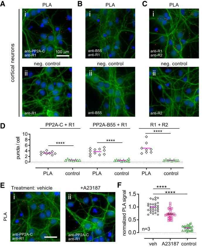
Submitted references Direct Interaction of PP2A Phosphatase with GABA(B) Receptors Alters Functional Signaling.
Li X, Terunuma M, Deeb TG, Wiseman S, Pangalos MN, Nairn AC, Moss SJ, Slesinger PA
The Journal of neuroscience : the official journal of the Society for Neuroscience 2020 Apr 1;40(14):2808-2816
The Journal of neuroscience : the official journal of the Society for Neuroscience 2020 Apr 1;40(14):2808-2816
No comments: Submit comment
Supportive validation
- Submitted by
- Invitrogen Antibodies (provider)
- Main image

- Experimental details
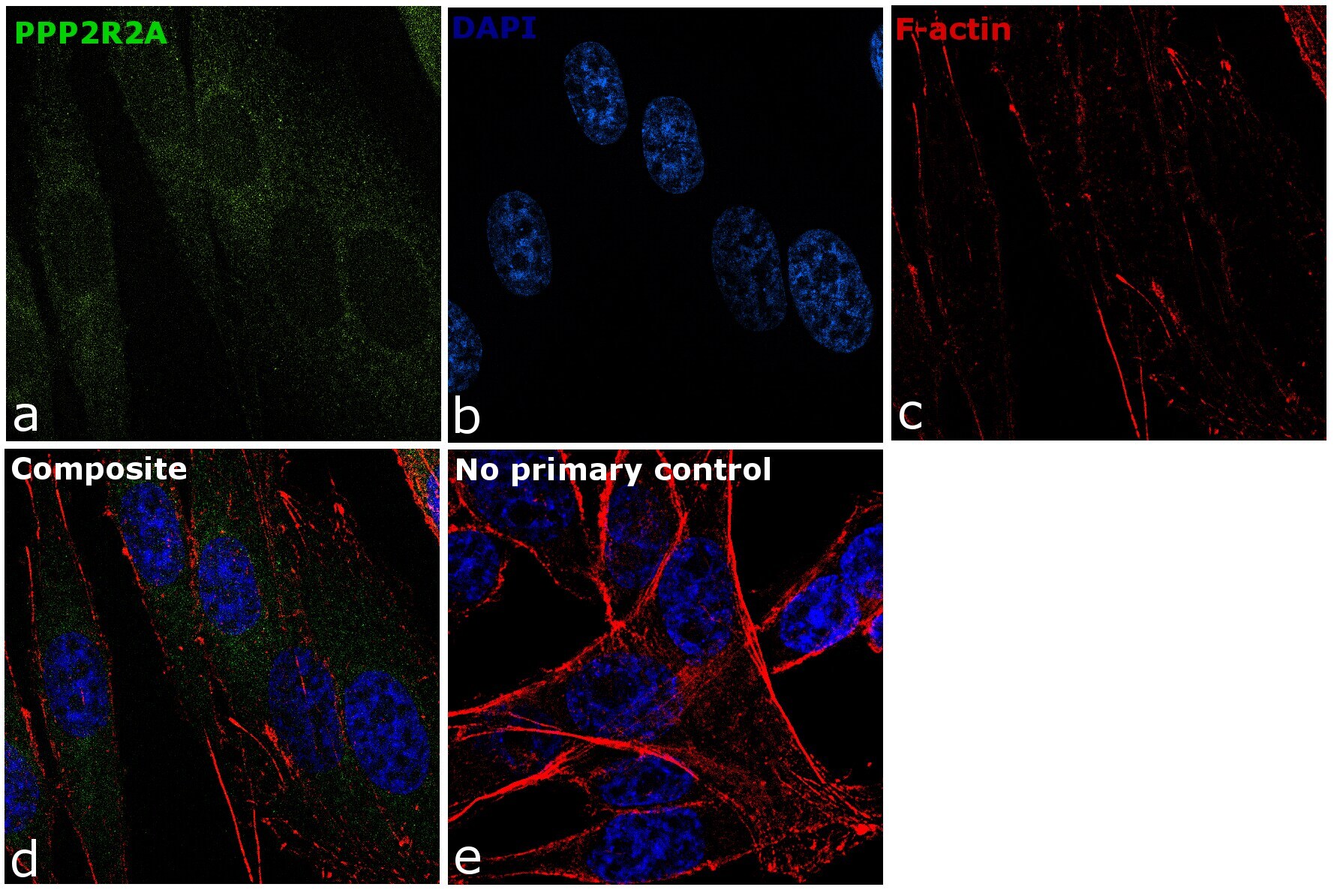
- Immunofluorescence analysis of PPP2R2A was performed using 70% confluent log phase PC-3 cells. The cells were fixed with 4% paraformaldehyde for 10 minutes, permeabilized with 0.1% Triton™ X-100 for 10 minutes, and blocked with 1% BSA for 1 hour at room temperature. The cells were labeled with PPP2R2A Monoclonal Antibody (Product # MA5-15007) at 5 µg/mL in 0.1% BSA and incubated overnight at 4 degree and then labeled with Goat anti-Rabbit IgG (H+L) Superclonal™ Secondary Antibody, Alexa Fluor® 488 conjugate (Product # A27034) at a dilution of 1:2000 for 45 minutes at room temperature (Panel a: green). Nuclei (Panel b: blue) were stained with SlowFade® Gold Antifade Mountant with DAPI (Product # S36938). F-actin (Panel c: red) was stained with Rhodamine Phalloidin (Product # R415, 1:300). Panel d represents the merged image showing cytoplasmic and nuclear localization. Panel e represents control cells with no primary antibody to assess background. The images were captured at 60X magnification.
- Submitted by
- Invitrogen Antibodies (provider)
- Main image

- Experimental details
- Immunofluorescence analysis of PPP2R2A was performed using 70% confluent log phase PC-3 cells. The cells were fixed with 4% paraformaldehyde for 10 minutes, permeabilized with 0.1% Triton™ X-100 for 10 minutes, and blocked with 1% BSA for 1 hour at room temperature. The cells were labeled with PPP2R2A Monoclonal Antibody (Product # MA5-15007) at 5 µg/mL in 0.1% BSA and incubated overnight at 4 degree and then labeled with Goat anti-Rabbit IgG (Heavy Chain) Superclonal™ Secondary Antibody, Alexa Fluor® 488 conjugate (Product # A27034) at a dilution of 1:2000 for 45 minutes at room temperature (Panel a: green). Nuclei (Panel b: blue) were stained with SlowFade® Gold Antifade Mountant with DAPI (Product # S36938). F-actin (Panel c: red) was stained with Rhodamine Phalloidin (Product # R415, 1:300). Panel d represents the merged image showing cytoplasmic and nuclear localization. Panel e represents control cells with no primary antibody to assess background. The images were captured at 60X magnification.

Supportive validation
- Submitted by
- Invitrogen Antibodies (provider)
- Main image
- Experimental details
- NULL