Explore
 Validate
 Learn
Learn Western blot
Antibody data
- Antibody Data
- Antigen structure
- References [0]
- Comments [0]
- Validations
- Western blot [1]
- Immunocytochemistry [1]
- Immunoprecipitation [1]
- Immunohistochemistry [1]
Submit
Validation data
Reference
Comment
Report error
- Product number
- TA302077 - Provider product page

- Provider
- OriGene
- Proper citation
- OriGene Cat#TA302077, RRID:AB_2066775
- Product name
- Rabbit Polyclonal Antibody against BNIP3L
- Antibody type
- Polyclonal
- Description
- Rabbit Polyclonal Antibody against BNIP3L
- Host
- Rabbit
- Conjugate
- Unconjugated
- Epitope
- BNIP3L
- Antibody clone number
- NULL
- Vial size
- 100 µg
- Concentration
- 0.25 mg/ml
No comments: Submit comment
Supportive validation
- Submitted by
- OriGene (provider)
- Main image
- Experimental details
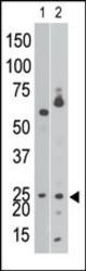
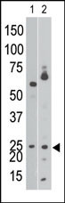
- The anti-NIP3 BH3 domain Pab (Cat. #TA302077) is used in Western blot to detect NIP3 BH3 in Ramos cell lysate (lane 1) and in mouse brain tissue lysate (lane 2).
- Validation comment
- WB
Supportive validation
- Submitted by
- OriGene (provider)
- Main image
- Experimental details
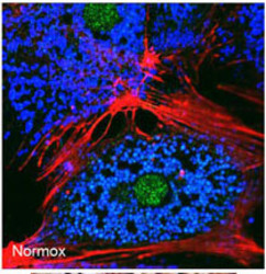
- Freshly isolated mouse hepatocytes plated on coverslips (2 x10⁵ cells/22-mm glass coverslip) were cultured under normoxic conditions for 6 hr. The cells were then fixed in 2% paraformaldehyde in PBS for 1 hr, and processed for confocal immunofluorescence (red: F-actin, blue: ATP-synthase, green: BNIP3). Fluorescence labeling of BNIP3 accomplished with anti-BNIP3 antibody Cat # TA302077. Data courtesy of Ruben Zamora, University of Pittsburgh.
- Validation comment
- IF

Supportive validation
- Submitted by
- OriGene (provider)
- Main image
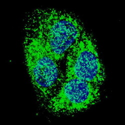
- Experimental details
- IF image of HepG2 cells stained with BNIP3 (BH3 Domain Specific) antibody. HepG2 cells were incubated with TA302077 BNIP3 (BH3 Domain Specific) primary antibody (1:500, 2 h at RT). For secondary antibody, Alexa Fluor? 488 conjugated donkey anti-rabbit antibody (green) was used (1:1000, 1h). Nuclei were counterstained with Hoechst 33342 (blue) . BNIP3 immunoreactivity is localized to the cytoplasm of HepG2 cells.
- Validation comment
- IP

Supportive validation
- Submitted by
- OriGene (provider)
- Main image

- Experimental details
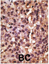
- Formalin-fixed and paraffin-embedded human cancer tissue reacted with the primary antibody, which was peroxidase-conjugated to the secondary antibody, followed by DAB staining. This data demonstrates the use of this antibody for immunohistochemistry; clinical relevance has not been evaluated. BC = breast carcinoma; HC = hepatocarcinoma.
- Validation comment
- IHC