Explore
 Validate
 Learn
Learn Western blot
 ELISA
ELISA Immunocytochemistry
Antibody data
- Antibody Data
- Antigen structure
- References [0]
- Comments [0]
- Validations
- Immunocytochemistry [2]
- Immunohistochemistry [2]
Submit
Validation data
Reference
Comment
Report error
- Product number
- MA5-35353 - Provider product page

- Provider
- Invitrogen Antibodies
- Product name
- GCN2 Recombinant Rabbit Monoclonal Antibody (1G2Z4)
- Antibody type
- Monoclonal
- Antigen
- Recombinant protein fragment
- Description
- Immunogen sequence: MAGGRGAPGR GRDEPPESYP QRQDHELQAL EAIYGADFQD LRPDACGPVK EPPEINLVLY PQGLTGEEVY VKVDLRVKCP PTYPDVVPEI ELKNAKGLSN ESVNLLKSRL EELAKKHCGE VMIFELAYHV QSFLSEHNKP PPKSFHEEML ERRAQEEQQR LLEAKRKEEQ EQREILHEIQ RRKEEIKEEK KRKEMAKQER LEIASLSNQD HTSKKDPGGH RTAAILHGGS PDFVGNGKHR ANSSGRSRRE RQYSVCNSED SPGSCEILYF NMGSPDQLMV HKGKCIGSDE QLGKLVYNAL
- Reactivity
- Human, Mouse, Rat
- Host
- Rabbit
- Isotype
- IgG
- Antibody clone number
- 1G2Z4
- Vial size
- 100 μL
- Concentration
- 1.0 mg/mL
- Storage
- -20°C, Avoid Freeze/Thaw Cycles
No comments: Submit comment
Supportive validation
- Submitted by
- Invitrogen Antibodies (provider)
- Main image
- Experimental details
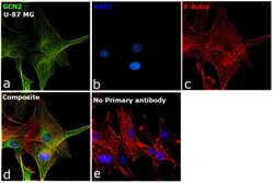
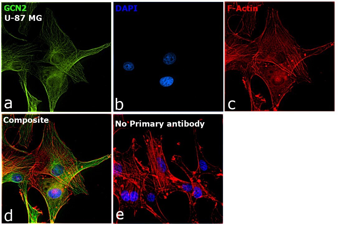
- Immunofluorescence analysis of GCN2 was performed using 70% confluent log phase U-87 MG cells. The cells were fixed with 4% paraformaldehyde for 10 minutes, permeabilized with 0.1% Triton™ X-100 for 15 minutes, and blocked with 2% BSA for 1 hour at room temperature. The cells were labeled with GCN2 Recombinant Rabbit Monoclonal Antibody (ARC0739) (Product # MA5-35353) at 1:200 dilution in 0.1% BSA, incubated at 4 degree celsius overnight and then labeled with Donkey anti-Rabbit IgG (H+L) Highly Cross-Adsorbed Secondary Antibody, Alexa Fluor Plus 488 (Product # A32790, 1:2000), for 45 minutes at room temperature (Panel a: Green). Nuclei (Panel b:Blue) were stained with ProLong™ Diamond Antifade Mountant with DAPI (Product # P36962). F-actin (Panel c: Red) was stained with Rhodamine Phalloidin (Product # R415, 1:300). Panel d represents the merged image showing Cytoskeleton and cytoplasmic localization. Panel e represents control cells with no primary antibody to assess background. The images were captured at 60X magnification.

- Submitted by
- Invitrogen Antibodies (provider)
- Main image

- Experimental details
- Immunofluorescence analysis of GCN2 was performed using 70% confluent log phase U-87 MG cells. The cells were fixed with 4% paraformaldehyde for 10 minutes, permeabilized with 0.1% Triton™ X-100 for 15 minutes, and blocked with 2% BSA for 1 hour at room temperature. The cells were labeled with GCN2 Recombinant Rabbit Monoclonal Antibody (ARC0739) (Product # MA5-35353) at 1:200 dilution in 0.1% BSA, incubated at 4 degree celsius overnight and then labeled with Donkey anti-Rabbit IgG (H+L) Highly Cross-Adsorbed Secondary Antibody, Alexa Fluor Plus 488 (Product # A32790, 1:2000), for 45 minutes at room temperature (Panel a: Green). Nuclei (Panel b:Blue) were stained with ProLong™ Diamond Antifade Mountant with DAPI (Product # P36962). F-actin (Panel c: Red) was stained with Rhodamine Phalloidin (Product # R415, 1:300). Panel d represents the merged image showing Cytoskeleton and cytoplasmic localization. Panel e represents control cells with no primary antibody to assess background. The images were captured at 60X magnification.
Supportive validation
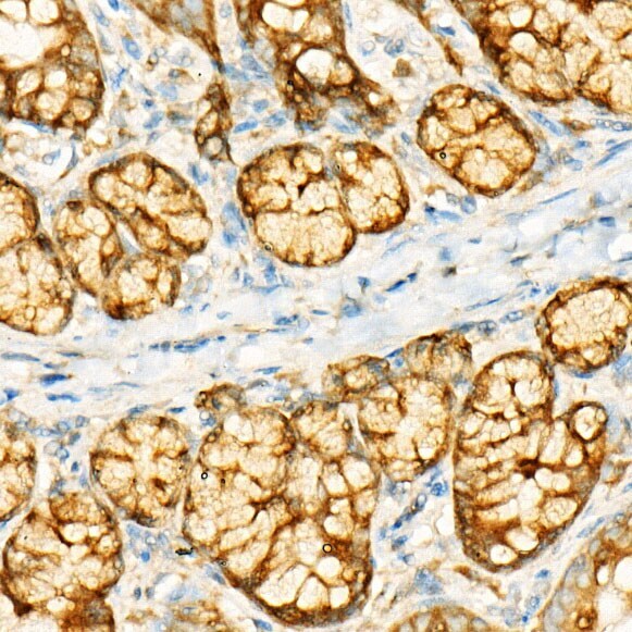
- Submitted by
- Invitrogen Antibodies (provider)
- Main image
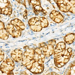
- Experimental details
- Immunohistochemistry analysis of GCN2 in paraffin-embedded rat Intestine. Samples were incubated with GCN2 Monoclonal antibody (Product # MA5-35353) using a dilution of 1:500 (40x lens). Perform high pressure antigen retrieval with 10 mM citrate buffer pH 6.0 before commencing with IHC staining protocol.
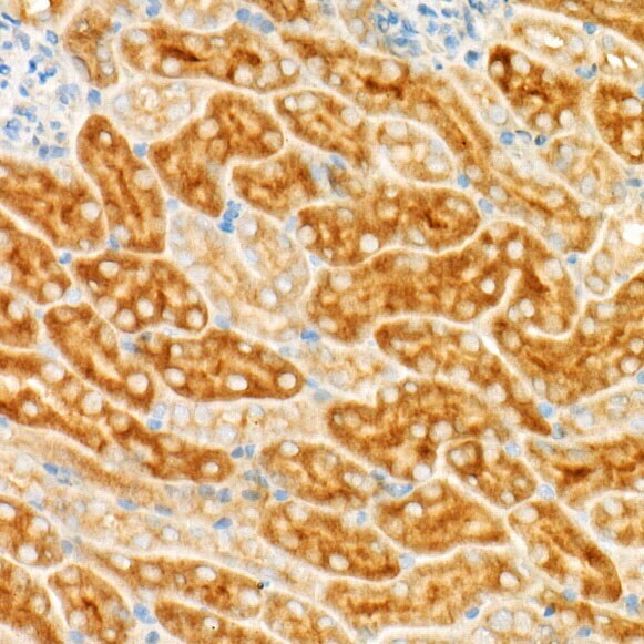
- Submitted by
- Invitrogen Antibodies (provider)
- Main image

- Experimental details
- Immunohistochemistry analysis of GCN2 in paraffin-embedded mouse kidney. Samples were incubated with GCN2 Monoclonal antibody (Product # MA5-35353) using a dilution of 1:500 (40x lens). Perform high pressure antigen retrieval with 10 mM citrate buffer pH 6.0 before commencing with IHC staining protocol.