Explore
 Validate
 Learn
Learn Western blot
 ELISA
ELISAAntibody data
- Antibody Data
- Antigen structure
- References [14]
- Comments [0]
- Validations
- Western blot [1]
- Immunohistochemistry [2]
- Flow cytometry [6]
Submit
Validation data
Reference
Comment
Report error
- Product number
- NB100-1869 - Provider product page

- Provider
- Novus Biologicals
- Proper citation
- Novus Cat#NB100-1869, RRID:AB_2190597
- Product name
- Rabbit Polyclonal EAAT1/GLAST-1/SLC1A3 Antibody
- Antibody type
- Polyclonal
- Description
- Immunogen affinity purified.
- Reactivity
- Human, Mouse, Rat
- Host
- Rabbit
- Isotype
- IgG
- Vial size
- 0.1 ml
- Concentration
- 1.0 mg/ml
- Storage
- Store at 4C short term. Aliquot and store at -20C long term. Avoid freeze-thaw cycles.
Submitted references Astrocytes have the capacity to act as antigen-presenting cells in the Parkinson's disease brain.
Single-Cell Transcriptomics Uncovers Glial Progenitor Diversity and Cell Fate Determinants during Development and Gliomagenesis.
Genetic ablation of Gpr37l1 delays tumor occurrence in Ptch1+/- mouse models of medulloblastoma.
Chronic hyperammonemia alters extracellular glutamate, glutamine and GABA and membrane expression of their transporters in rat cerebellum. Modulation by extracellular cGMP.
Depletion of microglia augments the dopaminergic neurotoxicity of MPTP.
Oct4 Methylation-Mediated Silencing As an Epigenetic Barrier Preventing Müller Glia Dedifferentiation in a Murine Model of Retinal Injury.
Decreased protein S-palmitoylation in dorsolateral prefrontal cortex in schizophrenia.
Exercise-Mediated Increase in Nigral Tyrosine Hydroxylase Is Accompanied by Increased Nigral GFR-α1 and EAAC1 Expression in Aging Rats.
Hypoxia-inducible factors enhance glutamate signaling in cancer cells.
Ceftriaxone increases glutamate uptake and reduces striatal tyrosine hydroxylase loss in 6-OHDA Parkinson's model.
Dopamine denervation of the prefrontal cortex increases expression of the astrocytic glutamate transporter GLT-1.
Transient striatal GLT-1 blockade increases EAAC1 expression, glutamate reuptake, and decreases tyrosine hydroxylase phosphorylation at ser(19).
Hypoxia regulates glutamate metabolism and membrane transport in rat PC12 cells.
Hypoxia regulates glutamate metabolism and membrane transport in rat PC12 cells.
Rostami J, Fotaki G, Sirois J, Mzezewa R, Bergström J, Essand M, Healy L, Erlandsson A
Journal of neuroinflammation 2020 Apr 16;17(1):119
Journal of neuroinflammation 2020 Apr 16;17(1):119
Single-Cell Transcriptomics Uncovers Glial Progenitor Diversity and Cell Fate Determinants during Development and Gliomagenesis.
Weng Q, Wang J, Wang J, He D, Cheng Z, Zhang F, Verma R, Xu L, Dong X, Liao Y, He X, Potter A, Zhang L, Zhao C, Xin M, Zhou Q, Aronow BJ, Blackshear PJ, Rich JN, He Q, Zhou W, Suvà ML, Waclaw RR, Potter SS, Yu G, Lu QR
Cell stem cell 2019 May 2;24(5):707-723.e8
Cell stem cell 2019 May 2;24(5):707-723.e8
Genetic ablation of Gpr37l1 delays tumor occurrence in Ptch1+/- mouse models of medulloblastoma.
Di Pietro C, La Sala G, Matteoni R, Marazziti D, Tocchini-Valentini GP
Experimental neurology 2019 Feb;312:33-42
Experimental neurology 2019 Feb;312:33-42
Chronic hyperammonemia alters extracellular glutamate, glutamine and GABA and membrane expression of their transporters in rat cerebellum. Modulation by extracellular cGMP.
Cabrera-Pastor A, Arenas YM, Taoro-Gonzalez L, Montoliu C, Felipo V
Neuropharmacology 2019 Dec 15;161:107496
Neuropharmacology 2019 Dec 15;161:107496
Depletion of microglia augments the dopaminergic neurotoxicity of MPTP.
Yang X, Ren H, Wood K, Li M, Qiu S, Shi FD, Ma C, Liu Q
FASEB journal : official publication of the Federation of American Societies for Experimental Biology 2018 Jun;32(6):3336-3345
FASEB journal : official publication of the Federation of American Societies for Experimental Biology 2018 Jun;32(6):3336-3345
Oct4 Methylation-Mediated Silencing As an Epigenetic Barrier Preventing Müller Glia Dedifferentiation in a Murine Model of Retinal Injury.
Reyes-Aguirre LI, Lamas M
Frontiers in neuroscience 2016;10:523
Frontiers in neuroscience 2016;10:523
Decreased protein S-palmitoylation in dorsolateral prefrontal cortex in schizophrenia.
Pinner AL, Tucholski J, Haroutunian V, McCullumsmith RE, Meador-Woodruff JH
Schizophrenia research 2016 Nov;177(1-3):78-87
Schizophrenia research 2016 Nov;177(1-3):78-87
Exercise-Mediated Increase in Nigral Tyrosine Hydroxylase Is Accompanied by Increased Nigral GFR-α1 and EAAC1 Expression in Aging Rats.
Arnold JC, Salvatore MF
ACS chemical neuroscience 2016 Feb 17;7(2):227-39
ACS chemical neuroscience 2016 Feb 17;7(2):227-39
Hypoxia-inducible factors enhance glutamate signaling in cancer cells.
Hu H, Takano N, Xiang L, Gilkes DM, Luo W, Semenza GL
Oncotarget 2014 Oct 15;5(19):8853-68
Oncotarget 2014 Oct 15;5(19):8853-68
Ceftriaxone increases glutamate uptake and reduces striatal tyrosine hydroxylase loss in 6-OHDA Parkinson's model.
Chotibut T, Davis RW, Arnold JC, Frenchek Z, Gurwara S, Bondada V, Geddes JW, Salvatore MF
Molecular neurobiology 2014 Jun;49(3):1282-92
Molecular neurobiology 2014 Jun;49(3):1282-92
Dopamine denervation of the prefrontal cortex increases expression of the astrocytic glutamate transporter GLT-1.
Vollbrecht PJ, Simmler LD, Blakely RD, Deutch AY
Journal of neurochemistry 2014 Jul;130(1):109-14
Journal of neurochemistry 2014 Jul;130(1):109-14
Transient striatal GLT-1 blockade increases EAAC1 expression, glutamate reuptake, and decreases tyrosine hydroxylase phosphorylation at ser(19).
Salvatore MF, Davis RW, Arnold JC, Chotibut T
Experimental neurology 2012 Apr;234(2):428-36
Experimental neurology 2012 Apr;234(2):428-36
Hypoxia regulates glutamate metabolism and membrane transport in rat PC12 cells.
Kobayashi S, Millhorn DE
Journal of neurochemistry 2001 Mar;76(6):1935-48
Journal of neurochemistry 2001 Mar;76(6):1935-48
Hypoxia regulates glutamate metabolism and membrane transport in rat PC12 cells.
Kobayashi S, Millhorn DE
Journal of neurochemistry 2001 Mar;76(6):1935-48
Journal of neurochemistry 2001 Mar;76(6):1935-48
No comments: Submit comment
Supportive validation
- Submitted by
- Novus Biologicals (provider)
- Main image

- Experimental details
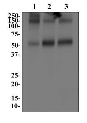
- Western Blot: EAAT1/GLAST-1/SLC1A3 Antibody [NB100-1869] - Analysis of SLC1A3 in 1. Human brain 2. Mouse brain and 3. Rat brain whole cell lysates.
Supportive validation
- Submitted by
- Novus Biologicals (provider)
- Main image
- Experimental details
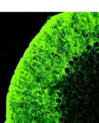
- Immunohistochemistry: EAAT1/GLAST-1/SLC1A3 Antibody [NB100-1869] - Staining of SLC1A3 in rat cerebellum sections

- Submitted by
- Novus Biologicals (provider)
- Main image

- Experimental details
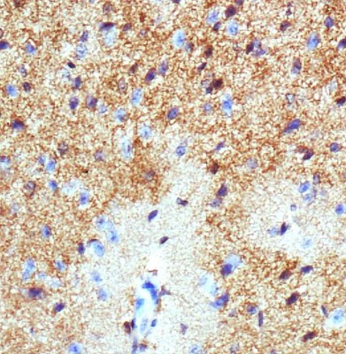
- Immunohistochemistry-Paraffin: EAAT1/GLAST-1/SLC1A3 Antibody [NB100-1869] - Analysis of SLC1A3 on mouse brain.
Supportive validation
- Submitted by
- Novus Biologicals (provider)
- Main image
- Experimental details
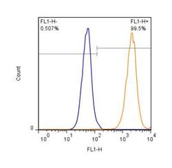
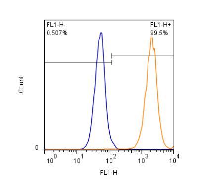
- Flow (Intracellular): EAAT1/GLAST-1/SLC1A3 Antibody [NB100-1869] - Intracellular staining of HEK293 cells (1 x 10^6 cells/mL) with SLC1A3 antibody (orange) stained at a dilution of 1:500. Detected with a GtxRb Dylight 488 secondary. Shown with the secondary control (blue).
- Submitted by
- Novus Biologicals (provider)
- Main image
- Experimental details
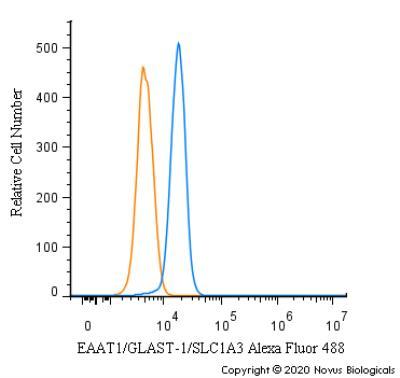
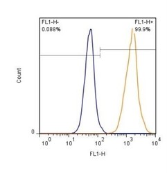
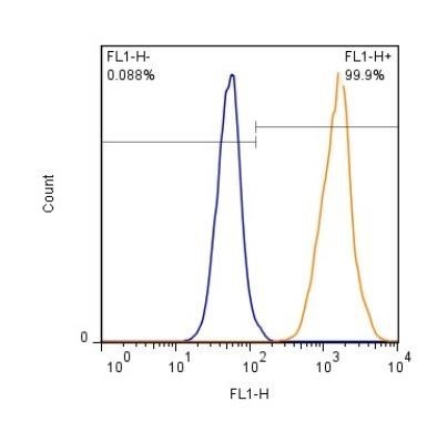
- Flow (Intracellular): EAAT1/GLAST-1/SLC1A3 Antibody [NB100-1869] - Staining of HEK293 cells (1 x 10^6 cells/mL) with AF488 conjugated EAAT-1 antibody (orange) stained at a dilution of 1:500. Shown with rIgG (AF488) isotype control (blue).
- Submitted by
- Novus Biologicals (provider)
- Main image
- Experimental details
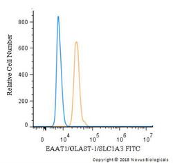
- Flow Cytometry: EAAT1/GLAST-1/SLC1A3 Antibody [NB100-1869] - An intracellular stain was performed on U-937 cells with NB100-1869F (blue) and a matched isotype control (orange). Cells were fixed with 4% PFA and then permeabilized with 0.1% saponin. Cells were incubated in an antibody dilution of 10 ug/mL for 30 minutes at room temperature. Both antibodies were conjugated to FITC.
- Submitted by
- Novus Biologicals (provider)
- Main image
- Experimental details
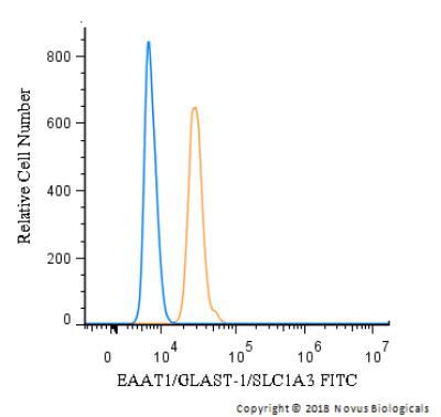
- Flow Cytometry: EAAT1/GLAST-1/SLC1A3 Antibody [NB100-1869] - An intracellular stain was performed on HeLa cells with NB100-1869F (blue) and a matched isotype control (orange). Cells were fixed with 4% PFA and then permeabilized with 0.1% saponin. Cells were incubated in an antibody dilution of 10 ug/mL for 30 minutes at room temperature. Both antibodies were conjugated to FITC.

- Submitted by
- Novus Biologicals (provider)
- Main image
- Experimental details
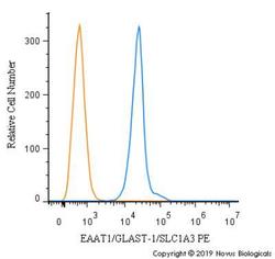
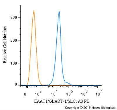
- Flow Cytometry: EAAT1/GLAST-1/SLC1A3 Antibody [NB100-1869] - An intracellular stain was performed on Hek293 cells with EAAT1/GLAST-1/SLC1A3 antibody NB100-1869PE (blue) and a matched isotype control (orange). Cells were fixed with 4% PFA and then permeablized with 0.1% saponin. Cells were incubated in an antibody dilution of 2.5 ug/mL for 30 minutes at room temperature. Both antibodies were conjugated to Phycoerythrin.
- Submitted by
- Novus Biologicals (provider)
- Main image
- Experimental details
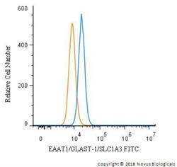
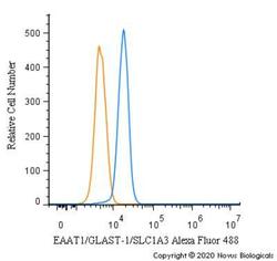
- Flow Cytometry: EAAT1/GLAST-1/SLC1A3 Antibody [NB100-1869] - An intracellular stain was performed on Hek293 cells with EAAT1/GLAST-1/SLC1A3 Antibody NB100-1869AF488 (blue) and a matched isotype control (orange). Cells were fixed with 4% PFA and then permeabilized with 0.1% saponin. Cells were incubated in an antibody dilution of 5 ug/mL for 30 minutes at room temperature. Both antibodies were conjugated to Alexa Fluor 488.