Explore
 Validate
 Learn
Learn Immunocytochemistry
Antibody data
- Antibody Data
- Antigen structure
- References [0]
- Comments [0]
- Validations
- Immunocytochemistry [1]
- Immunohistochemistry [1]
Submit
Validation data
Reference
Comment
Report error
- Product number
- HPA037467 - Provider product page

- Provider
- Atlas Antibodies
- Proper citation
- Atlas Antibodies Cat#HPA037467, RRID:AB_10795027
- Product name
- Anti-SLC1A3
- Antibody type
- Polyclonal
- Description
- Polyclonal Antibody against Human SLC1A3, Gene description: solute carrier family 1 (glial high affinity glutamate transporter), member 3, Alternative Gene Names: EA6, EAAT1, GLAST, Validated applications: ICC, IHC, Uniprot ID: P43003, Storage: Store at +4°C for short term storage. Long time storage is recommended at -20°C.
- Reactivity
- Human
- Host
- Rabbit
- Conjugate
- Unconjugated
- Isotype
- IgG
- Vial size
- 100 µl
- Concentration
- 0.1 mg/ml
- Storage
- Store at +4°C for short term storage. Long time storage is recommended at -20°C.
- Handling
- The antibody solution should be gently mixed before use.
No comments: Submit comment
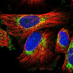
Supportive validation
- Submitted by
- Atlas Antibodies (provider)
- Main image
- Experimental details
- Immunofluorescent staining of human cell line U-251 MG shows localization to mitochondria.
- Sample type
- Human
Supportive validation
- Submitted by
- Atlas Antibodies (provider)
- Enhanced method
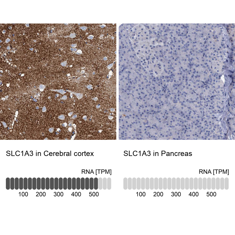
- Orthogonal validation
- Main image
- Experimental details
- Immunohistochemistry analysis in human cerebral cortex and pancreas tissues using HPA037467 antibody. Corresponding SLC1A3 RNA-seq data are presented for the same tissues.
- Sample type
- Human
- Protocol
- Protocol