Explore
 Validate
 Learn
Learn Western blot
 Immunocytochemistry
 Immunohistochemistry
Antibody data
- Antibody Data
- Antigen structure
- References [1]
- Comments [0]
- Validations
- Immunocytochemistry [1]
- Other assay [2]
Submit
Validation data
Reference
Comment
Report error
- Product number
- PA5-34601 - Provider product page

- Provider
- Invitrogen Antibodies
- Product name
- Stella Polyclonal Antibody
- Antibody type
- Polyclonal
- Antigen
- Synthetic peptide
- Description
- For Western Blot, this antibody has non-specific bands at 125 kDa and 25 kDa. This antibody is predicted to react with rat based on sequence homology.
- Reactivity
- Human, Mouse
- Host
- Rabbit
- Isotype
- IgG
- Vial size
- 100 μg
- Concentration
- 1 mg/mL
- Storage
- Store at 4°C short term. For long term storage, store at -20°C, avoiding freeze/thaw cycles.
Submitted references Identification of primordial germ cell-like cells as liver metastasis initiating cells in mouse tumour models.
Liu C, Ma Z, Cai Z, Zhang F, Liu C, Chen T, Peng D, Xu X, Lin HK
Cell discovery 2020;6:15
Cell discovery 2020;6:15
No comments: Submit comment
Supportive validation
- Submitted by
- Invitrogen Antibodies (provider)
- Main image
- Experimental details
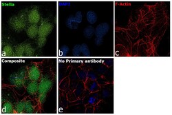
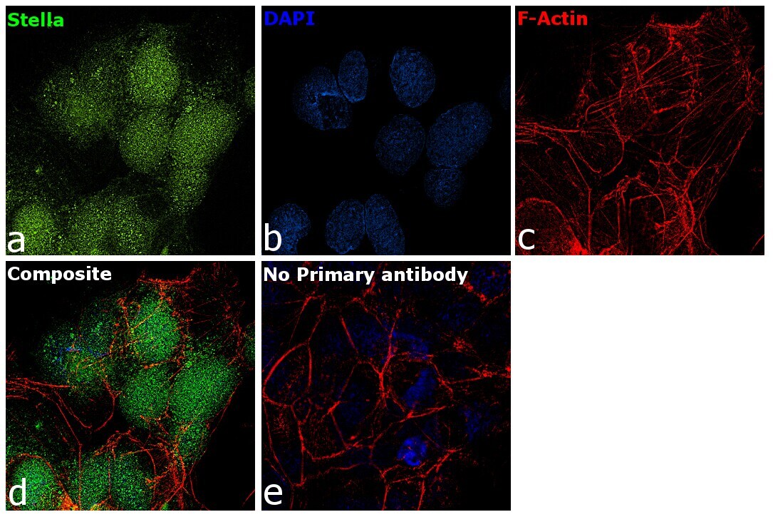
- Immunofluorescence analysis of Stella was performed using 70% confluent log phase BeWo cells. The cells were fixed with 4% paraformaldehyde for 10 minutes, permeabilized with 0.1% Triton™ X-100 for 15 minutes, and blocked with 2% BSA for 45 minutes at room temperature. The cells were labeled with Stella Polyclonal Antibody (Product # PA5-34601) at 5 µg/mL in 0.1% BSA, incubated at 4 degree celsius overnight and then labeled with Donkey anti-Rabbit IgG (H+L) Highly Cross-Adsorbed Secondary Antibody, Alexa Fluor Plus 488 (Product # A32790), (1:2000 dilution), for 45 minutes at room temperature (Panel a: Green). Nuclei (Panel b:Blue) were stained with ProLong™ Diamond Antifade Mountant with DAPI (Product # P36962). F-actin (Panel c: Red) was stained with Rhodamine Phalloidin (Product # R415, 1:300 dilution). Panel d represents the merged image showing nuclear localization. Panel e represents control cells with no primary antibody to assess background. The images were captured at 60X magnification.
Supportive validation
- Submitted by
- Invitrogen Antibodies (provider)
- Main image
- Experimental details
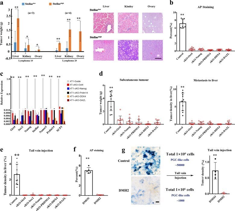
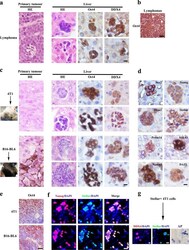
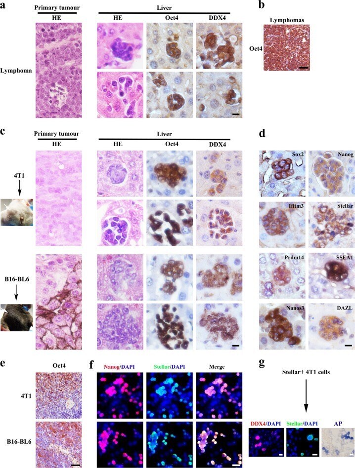
- Fig. 1 PGC-like subpopulation appears in early liver metastasis. a Primary lymphoma sections and liver tissues derived from p53 - / - mice were stained with H&E or primary antibody against indicated proteins. b Lymphoma sections from p53 - / - mice were stained with Oct4 antibody. c 4T1 breast cancer cells were injected into mammary fat pad of BALB/c mice, while B16-BL6 melanoma cells were injected subcutaneously into C57BL/6 mice. 4T1 and B16-BL6 primary tumors and their corresponding liver tissues were stained with H&E or the primary antibody against the indicated protein. d Representative sections of liver metastasis from 4T1 or B16-BL6 subcutaneous primary tumor were stained with the primary antibody against the indicated protein. e Primary Tumor sections from 4T1 and B16-BL6 cells were stained with antibody against indicated proteins. f The cultured 4T1 and B16-BL6 cells were stained with primary antibody against the indicated protein. g The fluorescent image of the daughter cells derived from Stellar high 4T1 cells 3 weeks after cultured. Scale bar = 20 mum in ( a , c , d , g ), 50 mum in ( b , e , f ).
- Submitted by
- Invitrogen Antibodies (provider)
- Main image

- Experimental details
- Fig. 6 Inhibition of PGC-like cell formation by PGC developmental pathways impairs liver metastasis. a The tumor weight and histological analysis of various tissues from the mice inoculated with Stellar low and Stellar high tumor cells sorted from two different lymphomas of p53 - / - mice through the tail vein injection. b The percentage of AP-positive cells in 4T1 cells with control and knockout in diverse PGC-related genes. c The real-time PCR analysis of the expression of diverse PGC-related genes in control and knockout 4T1 cells. d The weight of primary tumor and tumor density in liver from the mice subcutaneously injected with 4T1 cells with control and knockout in diverse PGC-related genes. e Tumor density in liver from the mice injected with 4T1 cells with control and knockout in diverse PGC-related genes 15 days after the tail vein injection. f The percentage of AP + cells in 4T1 cells treated with DMSO or DMH2 (6 muM). g Bright field image of AP staining of 4T1 cells treated with DMSO or DMH2 (6 muM). Tumor density in liver from the mice injected with 4T1 cells treated with DMSO or DMH2 for 6 days (6 muM). ** P < 0.01. Scale bar = 50 mum ( g ), 200 mum ( a ).