Explore
 Validate
 Learn
Learn Western blot
 Immunohistochemistry
Antibody data
- Antibody Data
- Antigen structure
- References [1]
- Comments [0]
- Validations
- Immunohistochemistry [2]
- Flow cytometry [2]
Submit
Validation data
Reference
Comment
Report error
- Product number
- AF1376 - Provider product page

- Provider
- R&D Systems
- Product name
- Human DLEC/CLEC4C/BDCA-2 Antibody
- Antibody type
- Polyclonal
- Description
- Antigen Affinity-purified. Detects human DLEC/CLEC4C/BDCA-2 in direct ELISAs and Western blots.
- Reactivity
- Human
- Host
- Goat
- Conjugate
- Unconjugated
- Antigen sequence
Q8WTT0- Isotype
- IgG
- Vial size
- 100 ug
- Storage
- Use a manual defrost freezer and avoid repeated freeze-thaw cycles. 12 months from date of receipt, -20 to -70 °C as supplied. 1 month, 2 to 8 °C under sterile conditions after reconstitution. 6 months, -20 to -70 °C under sterile conditions after reconstitution.
Submitted references In situ distribution of HIV-binding CCR5 and C-type lectin receptors in the human endocervical mucosa.
Hirbod T, Kaldensjö T, Broliden K
PloS one 2011;6(9):e25551
PloS one 2011;6(9):e25551
No comments: Submit comment
Supportive validation
- Submitted by
- R&D Systems (provider)
- Main image

- Experimental details
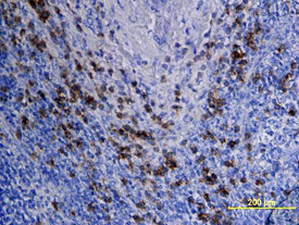
- DLEC/CLEC4C/BDCA-2 in Human Lymph Node. DLEC/CLEC4C/BDCA-2 was detected in immersion fixed paraffin-embedded sections of human lymph node using Goat Anti-Human DLEC/CLEC4C/BDCA-2 Antigen Affinity-purified Polyclonal Antibody (Catalog # AF1376) at 15 µg/mL overnight at 4 °C. Tissue was stained using the Anti-Goat HRP-DAB Cell & Tissue Staining Kit (brown; Catalog # CTS008) and counterstained with hematoxylin (blue). View our protocol for Chromogenic IHC Staining of Paraffin-embedded Tissue Sections.
- Submitted by
- R&D Systems (provider)
- Main image
- Experimental details
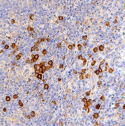
- DLEC/CLEC4C/BDCA-2 in Human Tonsil. DLEC/CLEC4C/BDCA-2 was detected in immersion fixed paraffin-embedded sections of human tonsil using Goat Anti-Human DLEC/CLEC4C/BDCA-2 Antigen Affinity-purified Polyclonal Antibody (Catalog # AF1376) at 3 µg/mL for 1 hour at room temperature followed by incubation with the Anti-Goat IgG VisUCyte™ HRP Polymer Antibody (Catalog # VC004). Tissue was stained using DAB (brown) and counterstained with hematoxylin (blue). Specific staining was localized to lymphocytes. View our protocol for IHC Staining with VisUCyte HRP Polymer Detection Reagents.

Supportive validation
- Submitted by
- R&D Systems (provider)
- Main image
- Experimental details
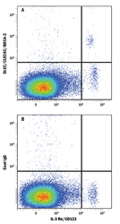
- Detection of DLEC/CLEC4C/BDCA-2 in Human PBMCs by Flow Cytometry. Human peripheral blood mononuclear cells (PBMCs) were stained with Mouse Anti-Human IL-3 R alpha/CD123 APC-conjugated Monoclonal Antibody (Catalog # FAB301A) and either (A) Goat Anti-Human DLEC/CLEC4C/BDCA-2 Antigen Affinity-purified Polyclonal Antibody (Catalog # AF1376) or (B) Normal Goat IgG Control (Catalog # AB-108-C) followed by Allophycocyanin-conjugated Anti-Goat IgG Secondary Antibody (Catalog # F0108).

- Submitted by
- R&D Systems (provider)
- Main image
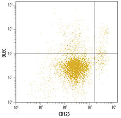
- Experimental details
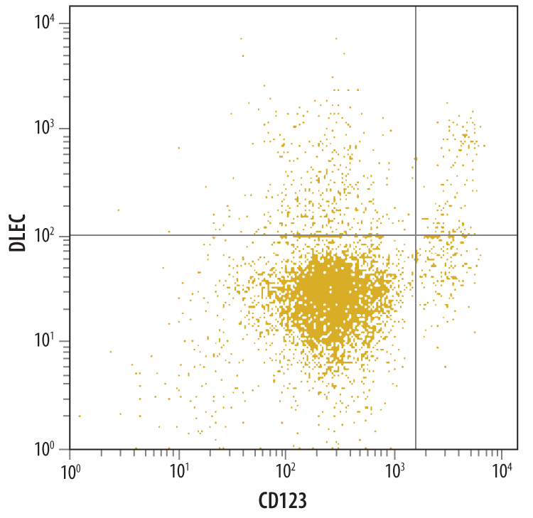
- Detection of DLEC/CLEC4C/BDCA-2 in Human Whole Blood by Flow Cytometry. Human whole blood were stained with Goat Anti- Human DLEC/CLEC4C/BDCA-2 Antigen Affinity-purified Polyclonal Antibody (Catalog # AF1376) followed by Allophycocyanin-conjugated Anti-Goat IgG Secondary Antibody (Catalog # F0108) and Human IL-3 R alpha Phycoerythrin-conjugated Monoclonal Antibody (Catalog # FAB301P).Quadrant markers were set based on control antibody staining (Catalog # AB-108-C).