 Explore
 Validate
 Learn
Learn Western blot
 Immunocytochemistry
Antibody data
- Antibody Data
- Antigen structure
- References [7]
- Comments [0]
- Validations
- Western blot [1]
- Immunohistochemistry [2]
Submit
Validation data
Reference
Comment
Report error
- Product number
- NBP1-22444 - Provider product page

- Provider
- Novus Biologicals
- Proper citation
- Novus Cat#NBP1-22444, RRID:AB_1659986
- Product name
- Mouse Monoclonal Cav3.2 Antibody
- Antibody type
- Monoclonal
- Description
- Protein G purified. Detects approx 260kDa. No cross-reactivity against Cav1.3.
- Reactivity
- Human, Mouse, Rat
- Host
- Mouse
- Isotype
- IgG
- Vial size
- 0.1 mg
- Concentration
- 1 mg/ml
- Storage
- Store at 4C short term. Aliquot and store at -20C long term. Avoid freeze-thaw cycles.
Submitted references Age attenuates the T-type Ca(V) 3.2-RyR axis in vascular smooth muscle.
Brain-derived neurotrophic factor stimulation of T-type Ca2+ channels in sensory neurons contributes to increased peripheral pain sensitivity.
Cav3.2 calcium channel interactions with the epithelial sodium channel ENaC.
Caveolae Link CaV3.2 Channels to BKCa-Mediated Feedback in Vascular Smooth Muscle.
T-type calcium channels functionally interact with spectrin (α/β) and ankyrin B.
TRPV1 Nociceptor Activity Initiates USP5/T-type Channel-Mediated Plasticity.
The deubiquitinating enzyme USP5 modulates neuropathic and inflammatory pain by enhancing Cav3.2 channel activity.
Fan G, Kaßmann M, Cui Y, Matthaeus C, Kunz S, Zhong C, Zhu S, Xie Y, Tsvetkov D, Daumke O, Huang Y, Gollasch M
Aging cell 2020 Apr;19(4):e13134
Aging cell 2020 Apr;19(4):e13134
Brain-derived neurotrophic factor stimulation of T-type Ca2+ channels in sensory neurons contributes to increased peripheral pain sensitivity.
Wang H, Wei Y, Pu Y, Jiang D, Jiang X, Zhang Y, Tao J
Science signaling 2019 Sep 24;12(600)
Science signaling 2019 Sep 24;12(600)
Cav3.2 calcium channel interactions with the epithelial sodium channel ENaC.
Garcia-Caballero A, Gandini MA, Huang S, Chen L, Souza IA, Dang YL, Stutts MJ, Zamponi GW
Molecular brain 2019 Feb 8;12(1):12
Molecular brain 2019 Feb 8;12(1):12
Caveolae Link CaV3.2 Channels to BKCa-Mediated Feedback in Vascular Smooth Muscle.
Hashad AM, Harraz OF, Brett SE, Romero M, Kassmann M, Puglisi JL, Wilson SM, Gollasch M, Welsh DG
Arteriosclerosis, thrombosis, and vascular biology 2018 Oct;38(10):2371-2381
Arteriosclerosis, thrombosis, and vascular biology 2018 Oct;38(10):2371-2381
T-type calcium channels functionally interact with spectrin (α/β) and ankyrin B.
Garcia-Caballero A, Zhang FX, Hodgkinson V, Huang J, Chen L, Souza IA, Cain S, Kass J, Alles S, Snutch TP, Zamponi GW
Molecular brain 2018 May 2;11(1):24
Molecular brain 2018 May 2;11(1):24
TRPV1 Nociceptor Activity Initiates USP5/T-type Channel-Mediated Plasticity.
Stemkowski P, García-Caballero A, Gadotti VM, M'Dahoma S, Huang S, Black SAG, Chen L, Souza IA, Zhang Z, Zamponi GW
Cell reports 2016 Dec 13;17(11):2901-2912
Cell reports 2016 Dec 13;17(11):2901-2912
The deubiquitinating enzyme USP5 modulates neuropathic and inflammatory pain by enhancing Cav3.2 channel activity.
García-Caballero A, Gadotti VM, Stemkowski P, Weiss N, Souza IA, Hodgkinson V, Bladen C, Chen L, Hamid J, Pizzoccaro A, Deage M, François A, Bourinet E, Zamponi GW
Neuron 2014 Sep 3;83(5):1144-58
Neuron 2014 Sep 3;83(5):1144-58
No comments: Submit comment
Supportive validation
- Submitted by
- Novus Biologicals (provider)
- Main image
- Experimental details
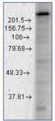
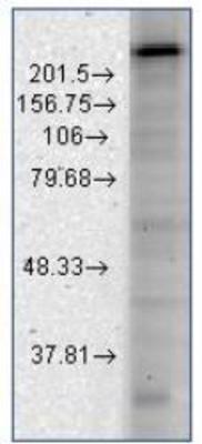
- Western Blot: Cav3.2 Antibody (S55-10) [NBP1-22444] - Western Blot using rat brain membranes and NBP1-22444 to target Cav3.2 channels visible at about 260kD.
Supportive validation
- Submitted by
- Novus Biologicals (provider)
- Main image
- Experimental details
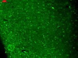
- Immunohistochemistry-Paraffin: Cav3.2 Antibody (S55-10) [NBP1-22444] - Tissue: hippocampus. Species: Human. Fixation: Bouin's Fixative and paraffin-embedded. Primary Antibody: Mouse Anti-CaV3.2 Calcium Channel Monoclonal Antibody at 1:1000 for 1 hour at RT. Secondary Antibody: FITC Goat Anti-Mouse (green) at 1:50 for 1 hour at RT.
- Submitted by
- Novus Biologicals (provider)
- Main image
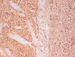
- Experimental details
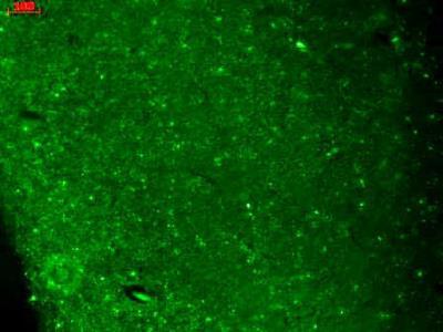
- Immunohistochemistry-Frozen: Cav3.2 Antibody (S55-10) [NBP1-22444] - Tissue: frozen brain section. Species: human. Fixation: 10% Formalin Solution for 12-24 hours at RT. Primary Antibody: Mouse Anti-CaV3.2 Calcium channel Monoclonal Antibody at 1:1000 for 1 hour at RT. Secondary Antibody: HRP/DAB Detection System: Biotinylated Goat Anti-Mouse, Streptavidin Peroxidase, DAB Chromogen (brown) for 30 minutes at RT. Counterstain: Mayer Hematoxylin (purple/blue) nuclear stain at 250-500 ul for 5 minutes at RT.
