Explore
 Validate
 Learn
Learn Flow cytometry
Antibody data
- Antibody Data
- Antigen structure
- References [3]
- Comments [0]
- Validations
- Flow cytometry [1]
- Other assay [2]
Submit
Validation data
Reference
Comment
Report error
- Product number
- 12-1579-41 - Provider product page

- Provider
- Invitrogen Antibodies
- Product name
- CD157 Monoclonal Antibody (eBioSY11B5 (SY11B5)), PE, eBioscience™
- Antibody type
- Monoclonal
- Antigen
- Other
- Description
- Description: The eBioSY11B5 monoclonal antibody recognizes human CD157 (Mo5, BST-1). CD157 is a 42-45 kDa, GPI-anchored protein with structural and functional similarities with CD38. CD157 was initially cloned because of its expression on monocytes and macrophages, and was subsequently discovered to be the same protein named BST-1, discovered for its expression on bone marrow stromal cells and its ability to stimulate the proliferation of a mouse pre-B cell line. CD157 is a pleiotropic ectoenzyme and is thought to act independently as an enzyme and receptor. Similar to CD38, CD157 is involved in the metabolism of NAD+ and this activity may be involved in regulating intracellular Ca2+ levels. As a receptor, upon binding of its putative ligand, CD157 is thought to initiate a signal transduction cascade resulting in the phosphorylation of cytoplasmic proteins including focal adhesion kinase (FAK). The mechanism and functional significance of CD157-initiated signal transduction remain to be fully characterized. Applications Reported: This eBioSY11B5 (SY11B5) antibody has been reported for use in flow cytometric analysis. Applications Tested: This eBioSY11B5 (SY11B5) antibody has been pre-titrated and tested by flow cytometric analysis of normal human peripheral blood cells. This can be used at 5 µL (0.125 µg) per test. A test is defined as the amount (µg) of antibody that will stain a cell sample in a final volume of 100 µL. Cell number should be determined empirically but can range from 10^5 to 10^8 cells/test. Excitation: 488-561 nm; Emission: 578 nm; Laser: Blue Laser, Green Laser, Yellow-Green Laser. Filtration: 0.2 µm post-manufacturing filtered.
- Reactivity
- Human
- Host
- Mouse
- Conjugate
- Yellow dye
- Isotype
- IgG
- Antibody clone number
- eBioSY11B5 (SY11B5)
- Vial size
- 25 Tests
- Concentration
- 5 µL/Test
- Storage
- 4° C, store in dark, DO NOT FREEZE!
Submitted references Comparison of Human Antral Follicles of Xenograft versus Ovarian Origin Reveals Disparate Molecular Signatures.
CD157 Confers Host Resistance to Mycobacterium tuberculosis via TLR2-CD157-PKCzeta-Induced Reactive Oxygen Species Production.
Novel SCRG1/BST1 axis regulates self-renewal, migration, and osteogenic differentiation potential in mesenchymal stem cells.
Man L, Lustgarten-Guahmich N, Kallinos E, Redhead-Laconte Z, Liu S, Schattman B, Redmond D, Hancock K, Zaninovic N, Schattman G, Rosenwaks Z, James D
Cell reports 2020 Aug 11;32(6):108027
Cell reports 2020 Aug 11;32(6):108027
CD157 Confers Host Resistance to Mycobacterium tuberculosis via TLR2-CD157-PKCzeta-Induced Reactive Oxygen Species Production.
Yang Q, Liao M, Wang W, Zhang M, Chen Q, Guo J, Peng B, Huang J, Liu H, Yahagi A, Xu X, Ishihara K, Cooper A, Chen X, Cai Y
mBio 2019 Aug 27;10(4)
mBio 2019 Aug 27;10(4)
Novel SCRG1/BST1 axis regulates self-renewal, migration, and osteogenic differentiation potential in mesenchymal stem cells.
Aomatsu E, Takahashi N, Sawada S, Okubo N, Hasegawa T, Taira M, Miura H, Ishisaki A, Chosa N
Scientific reports 2014 Jan 13;4:3652
Scientific reports 2014 Jan 13;4:3652
No comments: Submit comment
Supportive validation
- Submitted by
- Invitrogen Antibodies (provider)
- Main image

- Experimental details
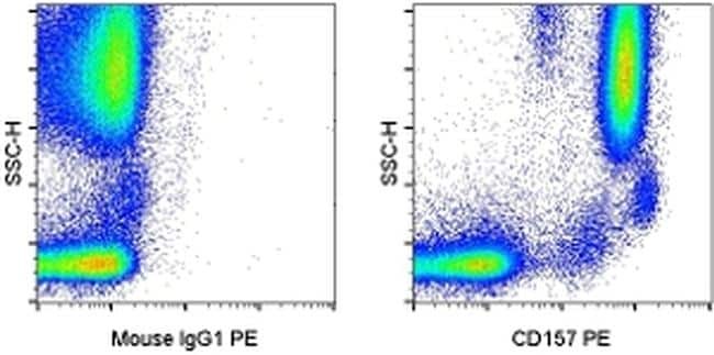
- Staining of normal human peripheral blood cells with Mouse IgG1 K Isotype Control PE (Product # 12-4714-81) (left) or Anti-Human CD157 PE (right).
- Conjugate
- Yellow dye
Supportive validation
- Submitted by
- Invitrogen Antibodies (provider)
- Main image
- Experimental details
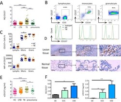
- FIG 1 CD157 expression levels are significantly increased in patients with TB. (A) Cd157 expression levels in whole blood from HC subjects ( n = 55), LTBI subjects ( n = 46), TB patients ( n = 51), and pneumonia patients ( n = 38) were determined by quantitative PCR (qPCR). RQ, relative quantification. (B) Peripheral blood was stained with anti-CD3, anti-CD14, and anti-CD157 antibodies and analyzed by flow cytometry. The histograms show the percentage of CD157-positive cells in lymphocytes, monocytes, and granulocytes from peripheral blood. SSC, side scatter. (C) The percentage (top panel) and mean fluorescence intensity (MFI) (bottom panel) of CD157 expression on lymphocytes (Lymphs), monocytes (Mons), and granulocytes (Grans) from HC subjects ( n = 21) and TB patients ( n = 20). (D) Immunohistochemical analysis of CD157 expression in tuberculous granuloma of lung tissue samples from patients with active tuberculosis (top panels) and in normal lung tissue adjacent to the lesions from patients with TB (bottom panels). Brown-labeled cells (arrow) represent CD157-positive cells. The images of the whole microscope slides were captured using a NanoZoomer digital pathology system (Hamamatsu Photonics). Outlined areas in the main images are enlarged in the insets. The bars represent 100 mum. Images that were representative of the images from three experiments were presented. (E) The concentrations of sCD157 in plasma from HC subjects ( n = 46), LTBI subjects ( n = 46), TB patients
- Conjugate
- Yellow dye
- Submitted by
- Invitrogen Antibodies (provider)
- Main image

- Experimental details
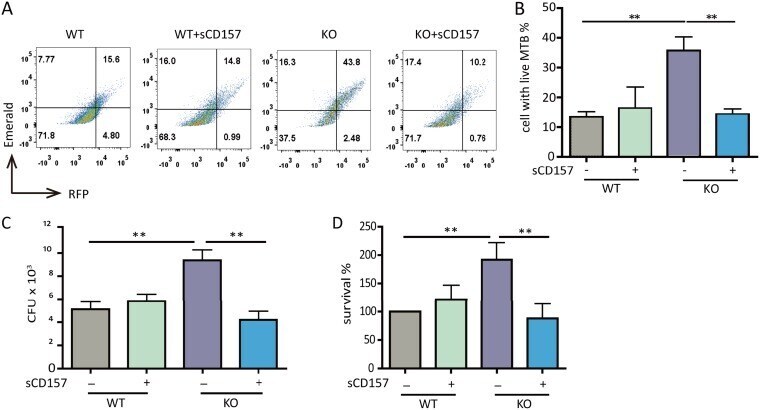
- FIG 3 CD157 deficiency impairs macrophage bactericidal capacity. Peritoneal macrophages from WT mice and Cd157 KO mice treated with sCD157 (+sCD157) (5 mug/ml) or not treated with sCD157 were infected with strain H37Ra harboring a dual-color reporter that comprises a constitutively green (Emerald) and a tetracycline-inducible red (TagRFP) fluorescent protein for 3 days. Tetracycline (500 ng/ml) was added 24 h before flow cytometry. Macrophages were then harvested and fixed with 4% PFA, and the percentage of cells with live M. tuberculosis (MTB) (red) were determined by FACS. The data are presented as representative dot plot data (A) and means plus SEM (B to D). Bacterial burden within peritoneal macrophages from WT mice and Cd157 KO mice at 72 h postinfection with the H37Rv strain (MOI of 5) in the presence or absence of sCD157 (5 mug/ml). The data were presented as absolute number of CFU (C) or the percentage of survival relative to WT mock treatment (D). One-way ANOVA Newman-Keuls multiple comparison test (B, C, and D) was used. * * , P
- Conjugate
- Yellow dye